

Review
doi: 10.1042/BC20040098.
Epidermal stem cells: the cradle of epidermal determination, differentiation and wound healing
Affiliations
- PMID: 15715523
- PMCID: PMC1283090
- DOI: 10.1042/BC20040098
Item in Clipboard
Review
Epidermal stem cells: the cradle of epidermal determination, differentiation and wound healing
Biol Cell.
2005 Mar.
Abstract
The field of epidermal stem cells has dramatically advanced in the last decade, leading to a better understanding of the molecular factors, signalling pathways and cellular events that identify and characterize stem cells, thus revealing their immense potential for therapeutic use. Furthermore, multipotent epidermal stem cells present the major advantage of easy accessibility with the discovery of their specific location within the bulge of the hair follicle. This review focuses on the most recent findings on epidermal stem cells, and their potential role in initial epidermal commitment, differentiation and wound healing processes in the skin.
Figures
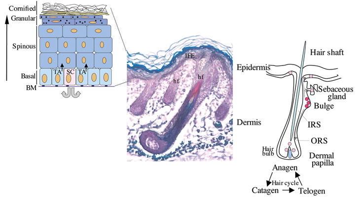
Left-hand panel, schematic representation of the stratified layers of the epidermis (basal, spinous, granular and cornified layers). The proliferative basal layer is adjacent to the basement membrane (BM). Stem cells (SC) generate transit amplifying (TA) cells which will differentiate to form the stratified layers. Middle panel, section of mouse skin stained to distinguish the different compartments of the hair follicle (hf) (Magnification 10×). Right-hand panel, schematic representation of a hair follicle and hair cycle, with the multipotent SCs (red) localizing to the bulge region. These cells migrate to populate the bulb region of the follicle, the sebaceous gland and the interfollicular epidermis (IFE; pink). IRS, inner root sheath; ORS, outer root sheath.
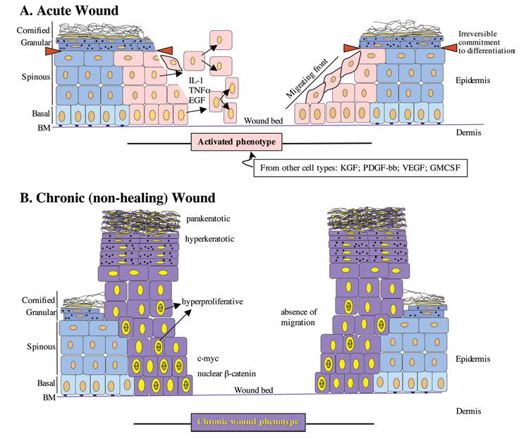
(A) In the acute wound, keratinocytes become activated (pink), and release/respond to various pro-inflammatory cytokines and growth factors. In response to these stimuli, keratinocytes from the basal and suprabasal layers start proliferating and migrating. Red triangles demarcate the point of irreversible commitment of keratinocytes to differentiation after which they are unable to participate in the wound healing response. (B) Chronic wound keratinocytes (purple) are hyperproliferative (indicated by mitotically active cells present throughout the suprabasal layers), hyperkeratotic (indicated by thick cornified layer) and parakeratotic (indicated by presence of nuclei in the cornified layer). Chronic wound keratinocytes are unable to migrate.
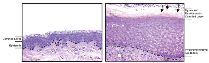
In the chronic ulcer, the epidermis is hyperproliferative, as indicated by increased thickness. The cornified layer is both thicker and contains cell nuclei (arrows). Dashed line: basement membrane. Magnification 20×.
References
-
- Abraham, J.A. and Klagsburn, M. (1996) Modulation of wound repair by members of the fibroblast growth factor family. In the molecular and cellular biology of wound repair (C.R.A., ed.), pp. 195–248, Plenum Press, New York
-
- Alonso L, Fuchs E. Stem cells in the skin: waste not, Wnt not. Genes Dev. 2003;17:1189–2000. - PubMed
-
- Arnold I, Watt FM. c-Myc activation in transgenic mouse epidermis results in mobilization of stem cells and differentiation of their progeny. Curr Biol. 2001;11:558–568. - PubMed
-
- Badavias EV, Abedi M, Butmarc J, Falanga V, Quesenberry P. Participation of bone marrow-derived cells in cutaneous wound healing. J Cell Physiol. 2003;196:245–250. - PubMed
-
- Banno T, Adachi M, Mukkamala L, Blumenberg M. Unique keratinocyte-specific effects of interferon-γ that protect skin from viruses identified using transcriptional profiling. Antiviral Ther. 2003;8:541–554. - PubMed
Publication types
MeSH terms
Grants and funding
LinkOut - more resources
Full Text Sources
Other Literature Sources
Medical
