. 2005 Feb 16;6(1):7.
doi: 10.1186/1471-2121-6-7.

Contribution of gap junctional communication between tumor cells and astroglia to the invasion of the brain parenchyma by human glioblastomas

Affiliations

Contribution of gap junctional communication between tumor cells and astroglia to the invasion of the brain parenchyma by human glioblastomas

Roxane Oliveira et al. BMC Cell Biol. .

Abstract

Background: Gliomas are "intraparenchymally metastatic" tumors, invading the brain in a non-destructive way that suggests cooperation between glioma cells and their environment. Recent studies using an engineered rodent C6 tumor cell line have pointed to mechanisms of invasion that involved gap junctional communication (GJC), with connexin 43 as a substrate. We explored whether this concept may have clinical relevance by analyzing the participation of GJC in human glioblastoma invasion.

Results: Three complementary in vitro assays were used: (i) seeding on collagen IV, to analyze homocellular interactions between tumor cells (ii) co-cultures with astrocytes, to study glioblastoma/astrocytes relationships and (iii) implantation into organotypic brain slice cultures, that mimic the three-dimensional parenchymal environment. Carbenoxolone, a potent blocker of GJC, inhibited cell migration in the two latter models. It paradoxically increased it in the first one. These results showed that homocellular interaction between tumor cells supports intercellular adhesion, whereas heterocellular glioblastoma/astrocytes interactions through functional GJC conversely support tumor cell migration. As demonstrated for the rodent cell line, connexin 43 may be responsible for this heterocellular functional coupling. Its levels of expression, high in astrocytes, correlated positively with invasiveness in biopsied tumors.

Conclusions: our results underscore the potential clinical relevance of the concept put forward by other authors based on experiments with a rodent cell line, that glioblastoma cells use astrocytes as a substrate for their migration by subverting communication through connexin 43-dependent gap junctions.


Figures

Figure 1
Figure 1
Heterocellular coupling of glioma cell lines, and migration assay in brain slice cultures. A, heterocellular coupling between astrocytes and human glioma cell lines (using the preloading method, see also fig. 6A). In contrast to 8-MG cells, GL15 cells established extensive GJC with normal astrocytes. Histograms represent mean heterocellular coupling indices (number of recipient astrocytes per double labeled tumor cell) of a minimum of three independent experiments (range 4 – 6; n= 120 to 225 donor cells per group; ± SEM; ***, p < 0.001). B, 8-MG cells and GL15 cells migration in brain slice cultures. Histograms of the average migration indices showing that GL15 were more invasive than 8-MG cells (mean values ± SEM. ***, p < 0.001).
Figure 2
Figure 2
Effects of inhibition of heterocellular coupling between GL15 cells and astrocytes. A, Photomicrographs in co-cultures (preloading method) showing heterocellular coupling without (upper panel) or with addition of the inhibitor CBX. GL15 donor cells pre-labeled with a non-diffusing membrane-bound dye (DiI, red) and loaded with a fluorescent gap junction-permeable dye (calcein, green) were seeded on a monolayer of unlabeled astrocytes. Functional heterocellular GJC is visualized as the transfer of calcein from DiI-labeled donor cells (small arrows) to surrounding recipients cells. Donors cells appear yellow because of the merge of red and green labeling. CBX reduced the number of calcein-containing (green) recipient astrocytes, (×200). B, Histograms representing mean values of a minimum of three independent runs as in A (range 4 – 6; n = 160 to 225 donor cells per group ± SEM). (***, p < 0.001). C and D, histograms of the migration indices of GL15 in brain slices without or with CBX, illustrating decreased migration in the treated group (mean values ± SEM. **, p < 0.01). E, Cumulative migration indices plotted against time in co-cultures where GL15 cell spheroids were plated upon an astrocyte monolayer, without or with CBX; (S - S0) / S0 values were measured every 12 h, for 48 h. Integrated areas under the two curves in arbitrary units were compared, p < 0.0001.
Figure 3
Figure 3
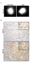
Inhibition of gap junctional communication in biopsied human gliomas. A. Vimentin immunostaining of the biopsied human glioblastoma HG-23 7 days after implantation into a cultured brain slice without (left panel) or with (right panel) CBX. Note the inhibition of cell migration when GJC is blocked. B, Connexin 43 immunocytochemistry (DAB, counterstained with hematoxylin) in two glioblastomas showing either moderate (HG-22) or abundant (HG-23) expression of the protein (×200). At the cellular level (inserts, ×1000), staining is dot-like or linear, predominantly at the membrane.
Figure 4
Figure 4
Connexin 43 expression in glioma cells. A, Western blot analysis of Cx43 expression in cell lines GL15 (lanes 1 and 2) and 8-MG (lanes 4 and 5), and in primary mouse astrocytes (lane 3). Three isoforms of Cx43 were detected in normal astrocytes, that corresponded to two phosphorylated (Cx43-P1 and Cx43-P2) and one non-phosphorylated (Cx43-NP) isoforms. GL15 cells exhibited higher level of Cx43 protein than 8 MG cells. B, Western blot analysis of Cx43 expression in seven human gliomas maintained in nude mice (T1 to T8). All tumors expressed one phosphorylated isoform. In addition, the non-phosphorylated isoform was detected in 4 tumor samples (T1, T2, T4 and T7). Tubulin was used as internal control The gels shown, are representative of a minimum of three independent experiments.
Figure 5
Figure 5
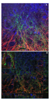
Cx43 immunoreactivity in brain slice culture injected with GL15 glioma cells. A, 3D projection of 15 consecutive confocal sections (0.5 μm) (×630), showing cooperation between migrating GL15 glioma cells (vimentin imunostaining, blue) with host astrocytes of the brain slice (GFAP immunostaining, red). Cx43 protein (green), is overexpressed by both tumor cells and astrocytes at the tumor margin where their processes are entwined. B, a single confocal section and two orthogonal projections (right and lower margins) corresponding to the overlayed perdendicular line. Enlarged view, of area boxed in B shows punctuate Cx43 immunoreactivity corresponding to gap junction plaques, at the interface of glioma cells and astrocytes strongly suggesting formation of heterocellular gap junctions. Note astrocyte morphologic changes which extend bipolar processes parallel to the long axes of the tumor cells
Figure 6
Figure 6
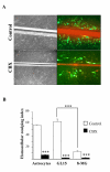
Homocellular coupling in human glioma cell lines using the scrape loading method. A, Dye coupling in absence (upper row) or presence (lower row) of the gap junctional blocker CBX. After scraping, injured cells were loaded with Lucifer Yellow (LY) and Rhodamin Dextran, and GJC was assessed by the number of neighboring cells that secondarily acquired green fluorescence. Left panel, confluent monolayer of GL15 cells in phase contrast. Right panel, centrifugal (left to right) diffusion of the dye tracer LY (green) from initially loaded injured cells (yellow, because of the merge of red and green) in the same monolayer. (×100). B, histogram representing the homocellular coupling indices (mean values ± SEM) from 4 to 6 independent cultures for each condition and cell type (astrocytes, GL15 or 8-MG). Homocellular coupling index = % labeled cells out of a total of ~400–800 analyzed cells per cell culture. Addition of CBX resulted in a dramatic decrease of the homocoupling in all cases ***, p < 0.001.
Figure 7
Figure 7
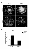
Migration assay of glioma cell lines on collagen IV. A, vimentin-immunostained GL15 and 8-MG spheroids, after 4 days of culture on collagen IV without (upper row) and with (lower row) addition of CBX, (x50). B, (S - S0) / S0 ratios (mean values ± SEM) obtained from 10 to 30 spheroids per group and condition. Addition of CBX enhanced spheroid disintegration and centrifugal dispersion of migrating cells. ***, p < 0.001.
Figure 8
Figure 8
In vivo parenchymal invasion of C6 glioma cells overexpressing the fusion protein GFP-Cx43. A, vimentin-immunolabeled C6 glioma cells (red) 8 days after striatal injection in the nude mice brain. C6 cells overexpressing the Cx43-GFP protein in green (arrow heads), are preferentially located at the tumor margin and display an invasive phenotype (×100). B, Microscopic field of the area boxed in A, at a higher magnificence (×200). C, D, confocal section (1 μm) of the border of another tumor showing invasive C6-Cx43-GFP cells at the tumor periphery migrating out. C6 cells which did not express Cx43-GFP protein, only labeled in red for vimentin remain in the tumor mass. E, is the overlay of the two channels. Migrating C6-Cx43-GFP cells appear yellow because of the merge of red and green. Dashed lines indicate the extremity of the tumor mass. Scale bar, 25 μm. tm, tumor mass.

References

    1. Bernstein JJ. Local invasion and intraparenchymal metastasis of astrocytomas. Neuropathol Appl Neurobiol. 1996;22:421–424. - PubMed
    1. Giese A, Westphal M. Glioma invasion in the central nervous system. Neurosurgery. 1996;39:235–50; discussion 250-2. doi: 10.1097/00006123-199608000-00001. - DOI - PubMed
    1. Reavey-Cantwell JF, Haroun RI, Zahurak M, Clatterbuck RE, Parker RJ, Mehta R, Fruehauf JP, Brem H. The prognostic value of tumor markers in patients with glioblastoma multiforme: analysis of 32 patients and review of the literature. J Neurooncol. 2001;55:195–204. doi: 10.1023/A:1013845004294. - DOI - PubMed
    1. Giese A, Kluwe L, Laube B, Meissner H, Berens ME, Westphal M. Migration of human glioma cells on myelin. Neurosurgery. 1996;38:755–764. doi: 10.1097/00006123-199604000-00026. - DOI - PubMed
    1. Giese A, Bjerkvig R, Berens ME, Westphal M. Cost of migration: invasion of malignant gliomas and implications for treatment. J Clin Oncol. 2003;21:1624–1636. doi: 10.1200/JCO.2003.05.063. - DOI - PubMed

Publication types