. 2005 May;16(5):2339-48.
doi: 10.1091/mbc.e05-01-0025. Epub 2005 Feb 16.

Plasticity of B cell receptor internalization upon conditional depletion of clathrin

Affiliations

Plasticity of B cell receptor internalization upon conditional depletion of clathrin

Angela Stoddart et al. Mol Biol Cell. 2005 May.

Abstract

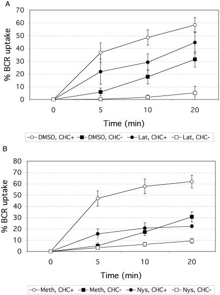
B cell antigen receptor (BCR) association with lipid rafts, the actin cytoskeleton, and clathrin-coated pits influences B cell signaling and antigen presentation. Although all three cellular structures have been separately implicated in BCR internalization, the relationship between them has not been clearly defined. In this study, internalization pathways were characterized by specifically blocking each potential mechanism of internalization. BCR uptake was reduced by approximately 70% in B cells conditionally deficient in clathrin heavy chain expression. Actin or raft antagonists were both able to block the residual, clathrin-independent BCR internalization. These agents also affected clathrin-dependent internalization, indicating that clathrin-coated pits, in concert with mechanisms dependent on rafts and actin, mediate the majority of BCR internalization. Clustering G(M1) gangliosides enhanced clathrin-independent BCR internalization, and this required actin. Thus, although rafts or actin independently did not mediate BCR internalization, they apparently cooperate to promote some internalization even in the absence of clathrin. Simultaneous inhibition of all BCR uptake pathways resulted in sustained tyrosine phosphorylation and activation of the extracellular signal-regulated kinase (ERK), strongly suggesting that downstream BCR signaling can occur without receptor translocation to endosomes and that internalization leads to signal attenuation.


Figures

Figure 1.
Figure 1.
Clathrin-mediated internalization in DKOS cells. (A) DKOS cells were treated with mouse anti-chicken IgM at 4°C, incubated at 37°C for 0 and 5 min, and then fixed and permeabilized. BCR was detected with an FITC-conjugated anti-mouse IgG and clathrin with polyclonal anti-CLC rabbit serum plus Rhodamine Red-conjugate anti-rabbit IgG. Immunofluorescent images of a central cell section showing the localization of the BCR (green), clathrin (red), and nucleus (blue). Yellow arrowheads denote colocalized BCR and CHC. Scale bar, 10 μM. (B) DKOS cells were untreated (-) or treated with 10 μg/ml goat anti-chicken IgM at 37°C for 0–20 min. CHC, immunoprecipitated with the mAb X22, was resolved by electrophoresis and immunoblotted for phosphotyrosine (pTyr) and CHC.
Figure 2.
Figure 2.
Conditional deletion of clathrin inhibits BCR internalization. DKOS cells were grown with (+) or without (−) 0.5 μg/ml doxycycline for 96 h. (A) Ubiquitous clathrin heavy chain (CHC), clathrin heavy chain isoform CHC22, and actin expression were determined by immunoblotting with anti-clathrin heavy chain mAb (TD.1), anti-CHC22 rabbit antiserum, and anti-actin mAb (AC-40). CHC** designates that this blot was overexposed to show residual CHC expression. (B) Cells were treated with 10 μg/ml goat anti-chicken IgM or biotinylated transferrin for 30 min at 4°C, washed, and warmed to 37°C for the times indicated. To detect receptors remaining on the cell surface, cells were stained on ice with FITC-labeled anti-goat Ig or Rhodamine Red-labeled streptavidin and analyzed by flow cytometry. The percent uptake represents the percentage of cells that no longer stain for surface BCR or TfR relative to staining at 4°C.
Figure 3.
Figure 3.
DKOS viability is not compromised by CHC depletion in conditions used for the internalization assays.(A) DKOS cells were plated at 2 × 10⁴ cells/ml with (CHC) or without (CHC+) doxycycline and maintained for 5 d. A small sample was removed at each time point, and live cells were counted by trypan blue exclusion. (B) DKOS cells were plated at 3 × 10⁵ cells/ml with or without doxycycline and split 10-fold at 48 h (condition used for internalization assays). Cells were fixed in 70% ethanol overnight at 4°C and then centrifuged and resuspended in propidium iodide/RNase A solution (Sigma 50 μg/ml and 5 mg/ml, respectively) at room temperature for >30 min. The percentage of cells in each phase of the cell cycle was assessed by flow cytometry.
Figure 4.
Figure 4.
Pharmacological agents that disrupt actin or rafts inhibit BCR internalization. DKOS cells were grown with (CHC) or without (CHC+) doxycycline. (A) Cells were treated for 30 min with 2 μM latrunculin B (Lat), stock dissolved in DMSO, or the equivalent concentration of DMSO alone. (B) Cells were treated for 30 min with 25 μg/ml nystatin (Nys) in 0.05% methanol (Meth) or 0.05% Meth alone. BCR internalization was monitored after each treatment as described in Figure 2.
Figure 5.
Figure 5.
Clustering GM1 gangliosides induces BCR internalization in clathrin-deficient DKOS cells. (A) DKOS cells were grown with or without doxycycline as indicated (CHC− and CHC+, respectively). Goat anti-chicken IgM was prebound to cells at 4°C. After a 30-min incubation, unbound antibody was removed by washing. Cells were placed at 37°C for 0 or 10 min to induce internalization. Cholera toxin B subunit (CTB)-Alexa 594 (red) was either prebound (b and d) with the anti-IgM and then cross-linked (37°C) or bound to cells after BCR cross-linking (a, c, and e) (4°C). Cells were permeabilized, fixed, and labeled with phalloidin Alexa 488 (green) or donkey anti-goat cy5 (blue) to stain actin and the BCR, respectively. (B) DKOS grown in doxycycline to deplete CHC were pretreated with DMSO or latrunculin B (Lat). CTB-Alexa 594 and anti-IgM were prebound on ice and then cross-linked at 37°C for 0 and 10 min. Cells were further processed as described in A. Scale bar, 10 μm.
Figure 6.
Figure 6.
BCR internalization is not inhibited in a variant subclone, DKOR, upon conditional depletion of CHC. DKOR cells were grown with (+) or without (−) 0.5 μg/ml doxycycline for 96 h. (A) Ubiquitous clathrin heavy chain (CHC) and actin expression were determined by immunoblotting with anti-clathrin heavy chain mAb (TD.1) and anti-actin mAb (AC-40) in DKOR cells. CHC** designates that this blot was overexposed to reveal residual CHC expression. (B) DKOR cells were treated with 10 μg/ml goat anti-chicken IgM or biotinylated conalbumin (white egg transferrin) for 30 min at 4°C, washed, and warmed to 37°C for the times indicated. To detect receptors remaining on the cell surface, cells were stained on ice with FITC-labeled anti-goat Ig or Rhodamine Red-labeled streptavidin and analyzed by flow cytometry. The percent uptake represents the percentage of cells that no longer stain for surface BCR or TfR.
Figure 7.
Figure 7.
Inhibition of BCR endocytosis does not block BCR-stimulated signaling. DKOS cells were grown with (CHC) or without (CHC+) doxycycline for 96 h and pretreated with DMSO or latrunculin B. B cells were either not stimulated (−) or treated with 10 μg/ml goat anti-chicken IgM at 37°C for 10 and 30 min. Ten micrograms of lysate was resolved by electrophoresis and immunoblotted for phosphotyrosine (pTyr), CHC, unphosphorylated and phosphorylated ERK1 and ERK2, and actin. Only the ERK2 isoform is detected in DKOS cells.

References

    1. Brodsky, F. M. (1985). Clathrin structure characterized with monoclonal antibodies. I. Analysis of multiple antigenic sites. J. Cell Biol. 101, 2047–2054. - PMC - PubMed
    1. Brodsky, F. M., Chen, C. Y., Knuehl, C., Towler, M. C., and Wakeham, D. E. (2001). Biological basket weaving: formation and function of clathrin-coated vesicles. Annu. Rev. Cell Dev. Biol. 17, 517–568. - PubMed
    1. Brown, B. K., and Song, W. (2001). The actin cytoskeleton is required for the trafficking of the B cell antigen receptor to the late endosomes. Traffic 2, 414–427. - PubMed
    1. Buerstedde, J. M., and Takeda, S. (1991). Increased ratio of targeted to random integration after transfection of chicken B cell lines. Cell 67, 179–188. - PubMed
    1. Chen, C-Y. and Brodsky, F. M. (2005). Huntingtin-interacting protein 1 (Hip 1) and Hip 1-related protein (Hip 1R) bind the conserved sequences of clathrin light chains and thereby influence clathrin assembly in vitro and actin distribution in vivo. J. Biol. Chem. 280, 6109–6117. - PubMed

Publication types

LinkOut - more resources