. 2005 Feb 14;168(4):537-44.
doi: 10.1083/jcb.200410006.

Temporal separation of replication and recombination requires the intra-S checkpoint

Affiliations

Temporal separation of replication and recombination requires the intra-S checkpoint

Peter Meister et al. J Cell Biol. .

Abstract

In response to DNA damage and replication pausing, eukaryotes activate checkpoint pathways that prevent genomic instability by coordinating cell cycle progression with DNA repair. The intra-S-phase checkpoint has been proposed to protect stalled replication forks from pathological rearrangements that could result from unscheduled recombination. On the other hand, recombination may be needed to cope with either stalled forks or double-strand breaks resulting from hydroxyurea treatment. We have exploited fission yeast to elucidate the relationship between replication fork stalling, loading of replication and recombination proteins onto DNA, and the intra-S checkpoint. Here, we show that a functional recombination machinery is not essential for recovery from replication fork arrest and instead can lead to nonfunctional fork structures. We find that Rad22-containing foci are rare in S-phase cells, but peak in G2 phase cells after a perturbed S phase. Importantly, we find that the intra-S checkpoint is necessary to avoid aberrant strand-exchange events during a hydroxyurea block.


Figures
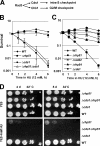
Figure 1.
Figure 1.
Functional recombination machinery is not essential for recovery from stalled replication forks. (A) Outline of checkpoint pathways in S. pombe. (B) Survival of recombination and S-phase checkpoint mutants after acute HU treatment. Isogenic cells of the confirmed genotypes were treated with 12 mM HU during the indicated times before plating in triplicate on YES medium. (C) Sensitivity of the indicated mutants in recombination pathways are compared with that of wild-type and cds1 cells after exposure to 12 mM HU for the indicated time and plating on solid YES medium for outgrowth. Experiments were performed at least twice and error bars are shown. (D) Sensitivity of the Δrhp51 cells to chronic HU exposure (4 mM).
Figure 2.
Figure 2.
Spatial separation of replication and recombination factories is affected by loss of the S-phase checkpoint. Low-level diffuse PCNA signals are typical of non-S-phase cells, whereas S-phase cells have a bright PCNA pattern. Although Rad22 foci are rare in wild-type cells, we show an example to illustrate the absence of colocalization with PCNA (enlarged to right). An example of colocalization is shown for cds1-deficient cells (enlarged in the right-most panel).
Figure 3.
Figure 3.
Hydroxyurea induces recombination foci in S-phase checkpoint mutants, not in wild-type or G2/M mutant cells. (A) Quantitation of the fraction of nuclei containing Rad22-YFP foci in asynchronously growing cells (−HU) or in cells treated with 12 mM HU for 2 h (+HU) in wild-type and checkpoint mutants. (B) Wild-type and Δcds1 strains were imaged either in the absence of HU or after a 2-h exposure to 12 mM HU. Left panels show Rad22-YFP foci are suppressed in cells with an intact S-phase checkpoint response during HU replication arrest, whereas the color image confirms that both cells have bright PCNA-CFP signals typical of S-phase cells.
Figure 4.
Figure 4.
Temporal separation of replication and recombination is affected by loss of the S-phase checkpoint. (A) The appearance of Rad22-YFP spots was monitored during an HU block and after release into fresh medium in wild-type and Δcds1 cells. Loss of the Cds1-mediated checkpoint leads to the accumulation of persistent Rad22 foci in S phase. In wild-type cells, Rad22 foci do not accumulate until release from HU arrest, and then rapidly disappear. This suggests a temporal separation of replication and recombination ensured by the S-phase checkpoint. Experiments were performed twice and error bars are shown. (B) Live imaging of ECFP-PCNA (green) and Rad22-YFP (red) in cells 30 min after release from HU arrest. Note that wild-type cells have few replication (PCNA) factories remaining when Rad22 foci appear, whereas the S-phase pattern persists in checkpoint-deficient cells, where PCNA and Rad22 foci often colocalize (white arrowheads). (C) Cds1 and Chk1 activation upon HU treatment and release in S-phase checkpoint or/and recombination mutants is monitored by Western blots. In each blot the top band represents the modified (activated) form of the kinase. (D) S-phase progression after release from HU treatment is restored by rhp51 deletion in checkpoint mutants. Cells deficient for S-phase checkpoint (Δcds1) are unable to resume replication after release from HU treatment, whereas strains deficient for both checkpoint and recombination can (Δcds1Δrhp51, black arrowhead).
Figure 5.
Figure 5.
rhp51 deletion partially rescues replication forks in intra-S checkpoint–deficient strain upon HU treatment. 2D gels analysis of replication intermediates at ars2.1 upon HU block and release in wild-type, Δcds1, Δrhp51, and Δcds1Δrhp51 cells are shown. Note that DNA replication intermediates were enriched by BND-cellulose chromatography, which is responsible for the low signal obtained in G2 cells (30 min after HU release) and the variable levels of 1n DNA (open arrowhead). In wild-type, Δrhp51, and Δcds1Δrhp51 cells replication intermediates (replication bubbles, black arrowhead; Y-forks, white arrowhead) persist upon HU treatment and disappear after release. In Δcds1 cells the bubble arc progressively disappears, and is replaced by double-Ys arcs and X-shaped forms (asterisk) that persist after HU release.

References

    1. Aparicio, O.M., A.M. Stout, and S.P. Bell. 1999. Differential assembly of Cdc45p and DNA polymerases at early and late origins of DNA replication. Proc. Natl. Acad. Sci. USA. 96:9130–9135. - PMC - PubMed
    1. Bjergbaek, L., Cobb, J.A., Tsai-Pflugfelder, M., and Gasser, S.M. 2005. Mechanistically distinct roles for Sgs1p in checkpoint activation and replication fork maintenance. EMBO J. 24:405–417. - PMC - PubMed
    1. Boddy, M.N., A. Lopez-Girona, P. Shanahan, H. Interthal, W.D. Heyer, and P. Russell. 2000. Damage tolerance protein Mus81 associates with the FHA1 domain of checkpoint kinase Cds1. Mol. Cell. Biol. 20:8758–8766. - PMC - PubMed
    1. Boddy, M.N., P. Shanahan, W.H. McDonald, A. Lopez-Girona, E. Noguchi, I.J. Yates, and P. Russell. 2003. Replication checkpoint kinase Cds1 regulates recombinational repair protein Rad60. Mol. Cell. Biol. 23:5939–5946. - PMC - PubMed
    1. Brewer, B.J., and W.L. Fangman. 1987. The localization of replication origins on ARS plasmids in S. cerevisiae. Cell. 51:463–471. - PubMed

Publication types

MeSH terms