. 2005 Feb 14;168(4):599-606.
doi: 10.1083/jcb.200409058.

Linking axonal degeneration to microtubule remodeling by Spastin-mediated microtubule severing

Affiliations

Linking axonal degeneration to microtubule remodeling by Spastin-mediated microtubule severing

Katia J Evans et al. J Cell Biol. .

Abstract

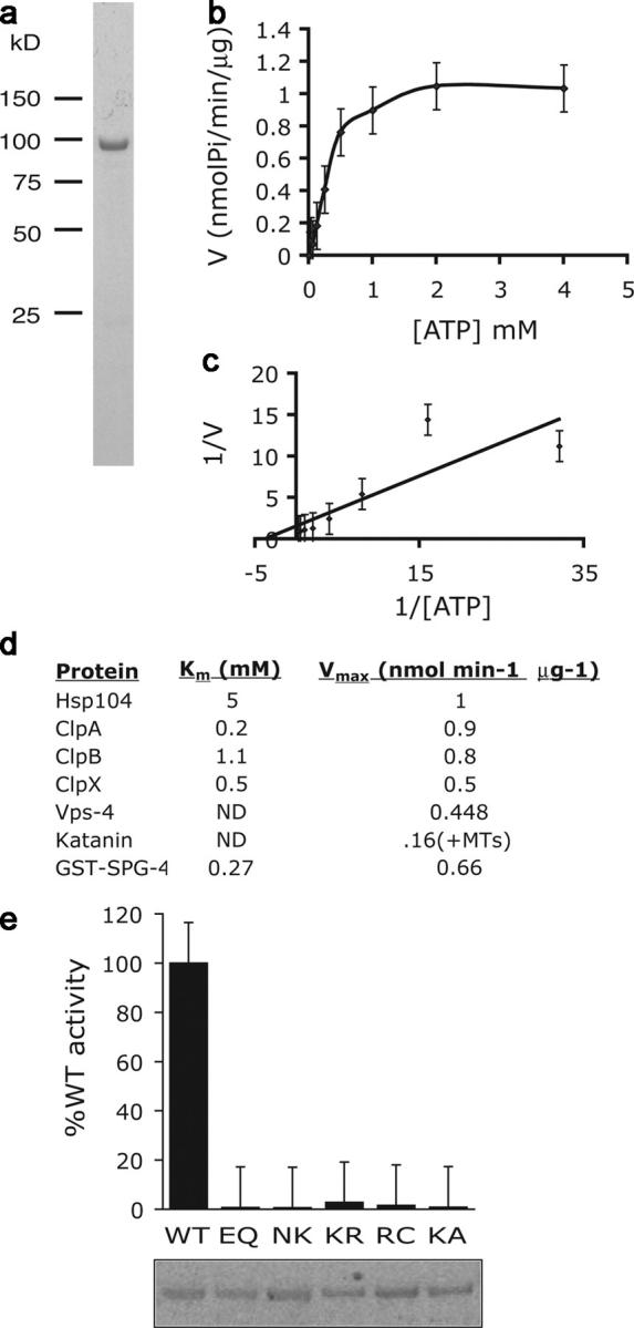
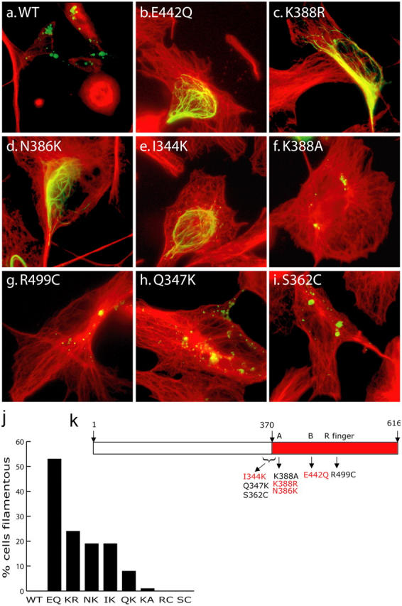
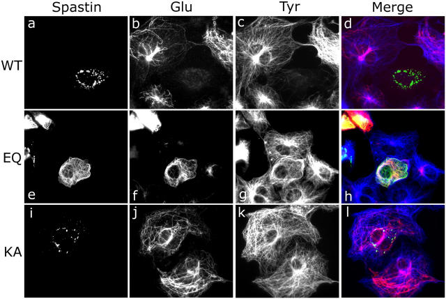
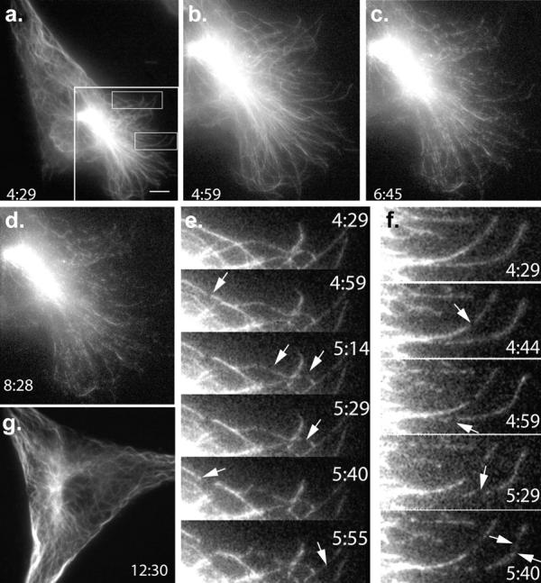
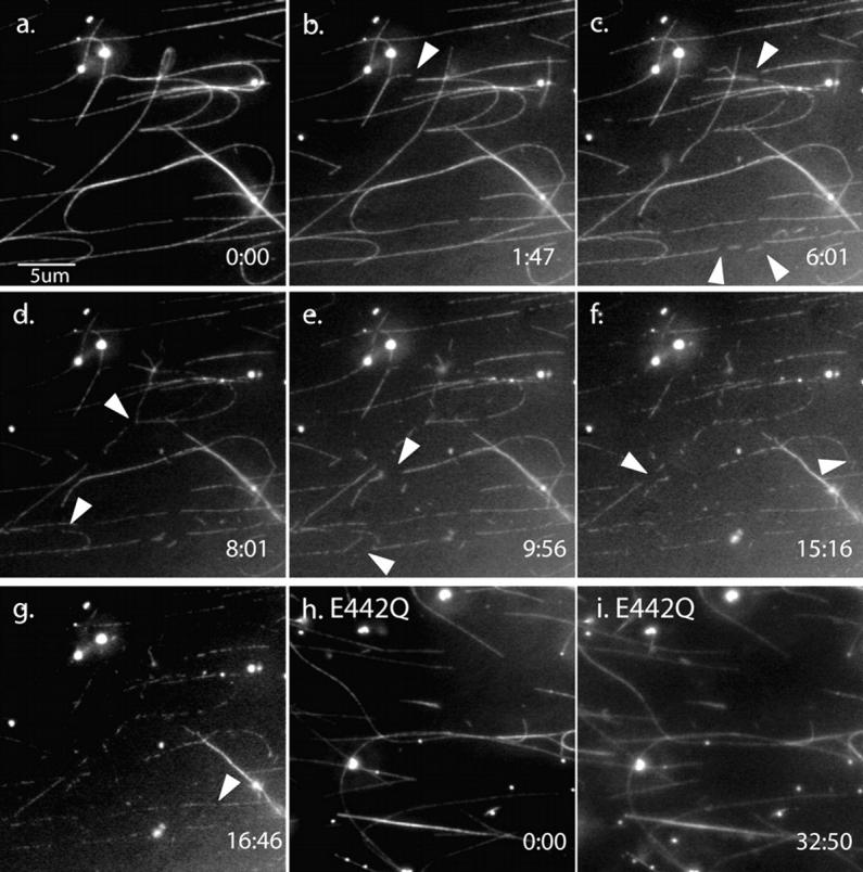
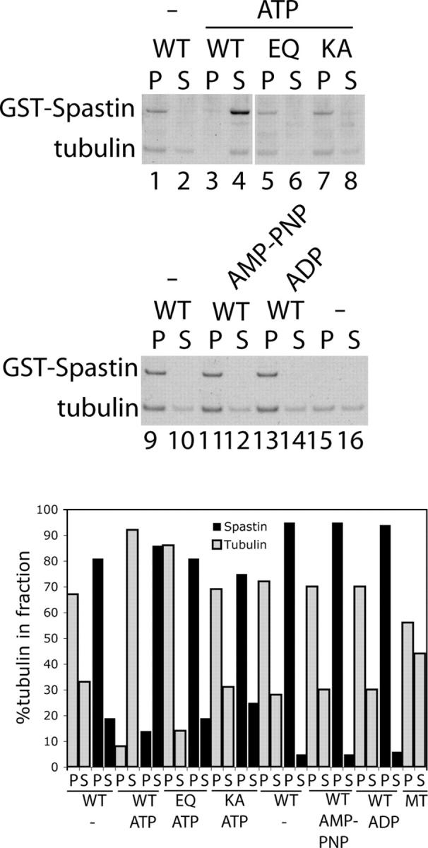
Mutations in the AAA adenosine triphosphatase (ATPase) Spastin (SPG4) cause an autosomal dominant form of hereditary spastic paraplegia, which is a retrograde axonopathy primarily characterized pathologically by the degeneration of long spinal neurons in the corticospinal tracts and the dorsal columns. Using recombinant Spastin, we find that six mutant forms of Spastin, including three disease-associated forms, are severely impaired in ATPase activity. In contrast to a mutation designed to prevent adenosine triphosphate (ATP) binding, an ATP hydrolysis-deficient Spastin mutant predicted to remain kinetically trapped on target proteins decorates microtubules in transfected cells. Analysis of disease-associated missense mutations shows that some more closely resemble the canonical hydrolysis mutant, whereas others resemble the ATP-binding mutant. Using real-time imaging, we show that Spastin severs microtubules when added to permeabilized, cytosol-depleted cells stably expressing GFP-tubulin. Using purified components, we also show that Spastin interacts directly with microtubules and is sufficient for severing. These studies suggest that defects in microtubule severing are a cause of axonal degeneration in human disease.


Figures

Figure 1.
Figure 1.
Analysis of recombinant Spastin. (a) 4 μg WT GST-Spastin was electrophoresed on a 12% gel. The fusion protein runs at ∼95 kD. (b) Saturation curve that shows ATPase activity of 15 nM WT GST-Spastin. 20-μl reactions were incubated for 20 min at 37°C. (c) Double reciprocal plot derived from the data in b is shown. (d) Km and Vmax were calculated from data in c and are shown in the table and compared with published values for other AAA ATPases; GST-SPG4 is WT Spastin (McNally and Vale, 1993; Babst et al., 1997; Schirmer et al., 1998). (e) Mutant versions of Spastin were prepared in parallel with WT enzyme and assayed for ATPase activity with 1 mM ATP for 15 min at 37°C with 25 nM enzyme. E442Q and K388A are discussed in the Results section. The other mutants represent disease-associated mutations. Note K388R and N386K are located in the Walker A P-loop, and that R499C is the arginine finger residue.
Figure 2.
Figure 2.
ATP hydrolysis deficient but not ATP binding deficient Spastin localizes to MTs. (a–i) TC 7 cells were transiently transfected with WT or mutant YFP-Spastin (green) for 24 h. Microtubule staining is in red. (a) WT Spastin forms cytosolic puncta, and transfected cells show decreased microtubule staining. (b) E442Q Spastin forms filaments that colocalize with a subset of microtubules. The percentage of transfected cells showing the filamentous pattern is shown in j.(f) K388A Spastin forms cytosolic puncta like the WT enzyme, however, the MT content of transfected cells is not decreased. Some disease-associated mutations (N386K, K388R, and I344K) decorated microtubules (c–e), whereas others did not (R499C, Q347K, and S362C) (g–i). (k) Schematic that shows the relative locations of the various mutations. Note two different mutations at the same position (K388A vs. K388R) produce different phenotypes.
Figure 3.
Figure 3.
Analysis of stable and dynamic microtubules in Spastin-expressing cells. Cos-7 cells were transiently transfected with YFP-Spastin constructs (green), WT (a–d), E442Q (e–h), or K388A (i–l) for 24 h. After methanol fixation, cells were stained for both Glu-tubulin (visualized with Cy-3–labeled secondary antibody; red) and Tyr-tubulin (visualized with Cy-5 labeled secondary antibody; blue). Compared with neighboring cells, WT Spastin overexpression results in decreased content of both stable and dynamic microtubules. Expression of the E442Q mutant shows that Spastin decorates microtubules and causes an increase in stable microtubules compared with nontransfected cells. Also, the microtubule organizing center appears less distinct in these cells. Expression of K388A Spastin does not appear to alter microtubule content or arrangement.
Figure 4.
Figure 4.
Spastin severs taxol-stabilized microtubules in permeabilized cells. (a–d) Extracted NIH 3T3 cells stably transfected with GFP-tubulin were permeabilized and microtubules were taxol stabilized before addition of WT-Spastin to 80 nM. After selecting a field to image, 0.25 mM ATP was added at time 0. Selected frames of the time-lapse are shown (min:s). (a) Whole-cell image (see Video 1, available at http://www.jcb.org/cgi/content/full/jcb.200409058/DC1). Panels b–d show a region delineated by the large box in panel a. In this case, microtubule severing is observed 5 min after the addition of ATP, and it occurs very rapidly (3 min). (e and f) Magnification of the two smaller boxed areas marked in panel a; arrows point to individual microtubules breaks (see Video 2). g. No severing occurs in a cell incubated with E442Q mutant and ATP even after 12 min (see Video 3). Bar, 10 μm.
Figure 5.
Figure 5.
Spastin severs purified microtubules in vitro. Rhodamine-labeled microtubules were assembled and immobilized in perfusion chambers containing ATP and an oxygen-scavenging system as described in Materials and methods. 100 nM WT GST-Spastin (a–g) or E442Q GST-Spastin in buffer containing ATP were perfused into the chambers, and images were acquired over a 20-min (WT incubation) or 32-min period (E442Q incubation). Selected frames are shown with the time (min:s) shown in each panel. Individual breaks in microtubules are first seen 1:47 min after addition of Spastin to the chamber (b) and continue to occur throughout 17 min (c–g). Arrowheads mark some of the new breaks observed in each frame. No breaks occur in incubations with ATP-hydrolysis deficient Spastin (h and i). The complete videos (Videos 4 and 5) are available at http://www.jcb.org/cgi/content/full/jcb.200409058/DC1. Some movement of microtubules is seen because of movement of liquid in the chamber, and some severed fragments are washed away.
Figure 6.
Figure 6.
Spastin is sufficient for severing MTs. Taxol-stabilized MTs were assembled from purified tubulin GTP and taxol and incubated with recombinant Spastin and nucleotides as indicated in the figure. After 10 min at 37°C, microtubules were separated from tubulin dimer by ultracentrifugation. In the absence of ATP, WT GST-Spastin sediments with microtubules (lanes 1 and 9), indicating direct binding. In contrast, with ATP, most of the tubulin was recovered in the supernatant fraction (lane 4), indicating that severing occurred. Note that Spastin does not sediment nonspecifically (lane 4). E442Q and K388A mutant Spastin sediment with microtubules but do not sever even in the presence of ATP (lanes 5 and 7). These results suggest that ATP hydrolysis is required for severing. Neither AMP-PNP nor ADP could substitute for ATP (lanes 11 and 13). The two panels represent separate experiments. (top gel) White line indicates that intervening lanes have been spliced out. Quantitation of the data from the gels is shown in the bar graph.

References

    1. Ahmad, F.J., W. Yu, F.J. McNally, and P.W. Baas. 1999. An essential role for katanin in severing microtubules in the neuron. J. Cell Biol. 145:305–315. - PMC - PubMed
    1. Antonsson, B., D.B. Kassel, G. Di Paolo, R. Lutjens, B.M. Riederer, and G. Grenningloh. 1998. Identification of in vitro phosphorylation sites in the growth cone protein SCG10. Effect of phosphorylation site mutants on microtubule-destabilizing activity. J. Biol. Chem. 273:8439–8446. - PubMed
    1. Babst, M., T.K. Sato, L.M. Banta, and S.D. Emr. 1997. Endosomal transport function in yeast requires a novel AAA-type ATPase, Vps4p. EMBO J. 16:1820–1831. - PMC - PubMed
    1. Babst, M., B. Wendland, E.J. Estepa, and S.D. Emr. 1998. The Vps4p AAA ATPase regulates membrane association of a Vps protein complex required for normal endosome function. EMBO J. 17:2982–2993. - PMC - PubMed
    1. Bhattacharya, R., and F. Cabral. 2004. A ubiquitous beta-tubulin disrupts microtubule assembly and inhibits cell proliferation. Mol. Biol. Cell. 15:3123–3131. - PMC - PubMed

Publication types