. 2005 Feb 16;25(7):1788-96.
doi: 10.1523/JNEUROSCI.4268-04.2005.

Toll-like receptor 4 on nonhematopoietic cells sustains CNS inflammation during endotoxemia, independent of systemic cytokines

Affiliations

Toll-like receptor 4 on nonhematopoietic cells sustains CNS inflammation during endotoxemia, independent of systemic cytokines

Sumana Chakravarty et al. J Neurosci. .

Abstract

Inflammatory agonists such as lipopolysaccharide (LPS) induce robust systemic as well as CNS responses after peripheral administration. Responses in the innate immune system require triggering of toll-like receptor 4 (TLR4), but the origin of CNS sequelas has been controversial. We demonstrate expression of TLR4 transcripts in mouse brain in the meninges, ventricular ependyma, circumventricular organs, along the vasculature, and in parenchymal microglia. The contribution of TLR4 expressed in CNS resident versus hematopoietic cells to the development of CNS inflammation was examined using chimeric mice. Reciprocal bone marrow chimeras between wild-type and TLR4 mutant mice show that TLR4 on CNS resident cells is critically required for sustained inflammation in the brain after systemic LPS administration. Hematopoietic TLR4 alone supported the systemic release of acute phase cytokines, but transcription of proinflammatory genes in the CNS was reduced in duration. In contrast, TLR4 function in radiation-resistant cells was sufficient for inflammatory progression in the brains of chimeric mice, despite the striking absence of cytokine elevations in serum. Surprisingly, a temporal rise in serum corticosterone was also dependent on TLR4 signaling in nonhematopoietic cells. Our findings demonstrate a requirement for TLR4 function in CNS-resident cells, independent of systemic cytokine effects, for sustained CNS-specific inflammation and corticosterone rise during endotoxemia.


Figures

Figure 1.
Figure 1.
Distribution of TLR4 transcripts in the mouse brain. A full-length 35S-labeled mouse TLR4 antisense riboprobe was hybridized to tissue sections singly (a-j) or in combination with antibodies (k-o) or a nonradioactive riboprobe (p-q). Dark-field image shows TLR4 mRNA expression in a spleen section from C3H/HeOuJ mice (a) and not in C57BL/10ScCr spleen (b). Insets are bright-field images. Dark-field image (d) corresponds to the brain area outlined in a Nissl-stained section (c). Circumventricular organs, SFO (e), ME (f), and AP (g) show TLR4 expression when viewed in dark field (e′-g′). Similarly, cells in choroid plexus (h, i), ventricular ependyma (j), and meninges (k) express TLR4 mRNA. Hybridization with radioactive TLR4 cRNA was followed by immunohistochemistry with NeuN (l, m) or a vascular marker isolectin B4 (k, n, o). Inset (k) shows a low-power dark-field view of TLR4 mRNA in the meninges over cortex (arrowhead). Radioactive TLR4 mRNA (silver grains in the dark-field image, p) overlapped with a nonradioactive CX3CR1 label (purple cells in p′). Magnified bright-field view (q) and inset showing one double-labeled cell. TLR4 mRNA also colocalized with Iba1-positive microglial cells (r, magnified in r′), but not with GFAP-labeled astrocytes (s). LV, Lateral ventricle; ChPlx, choroid plexus; PVN, paraventricular nucleus; 3V, third ventricle. Scale bars: a, b, d, 1 mm; e-h, 0.5 mm; i-o, 0.05 mm; p, q, r, 0.05 mm.
Figure 2.
Figure 2.
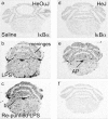
Effect of LPS repurification on gene transcription in TLR4 mutant mice. Film autoradiographs show induction of IκBα (a-f) in brainstem sections of mice 2 h after LPS administration. C3H/HeOuJ (a-c) or C3H/HeJ mice (d-f) were injected intraperitoneally with saline (a, d); LPS serotype 055:B5 (b, e) or repurified LPS (c, f). bv, blood vessel.
Figure 3.
Figure 3.
Hematopoietic reconstitution replaces >95% of resident CD45+ cells. We assessed the efficiency of hematopoietic chimerism in our experimental protocol by making MHC class I mismatched chimeras. Bone marrow from B6 mice (MHC haplotype b) or HeOuJ mice (MHC haplotype k) were transferred into irradiated HeJ mice (MHC haplotype k). Two months after reconstitution, spleens were isolated from chimeric mice. Single-cell suspensions from spleen were labeled with antibodies to CD45 as well as antibodies specific to the MHC Class I molecules (H-2K) of the k (H-2Kk) or b (H-2Kb) haplotype. Staining was analyzed on a BD FACScalibur. Plots show MHC staining observed on cells that are CD45+ (gated). In the case of mice receiving HeOuJ bone marrow (a), ∼96% of the CD45+ cells were of the HeOuJ origin. In the case of mice receiving the B6 bone marrow (b), ∼91% of the cells stained for the B6 Class I molecule. Although ∼2% of the cells fell within the bottom right quadrant, suggesting a staining by the k haplotype, there was no distinct population corresponding to the MHC-expressing cells (compare bottom right quadrants in a). This allows us to conclude that our protocols were successful in generating at least 90-95% donor-derived hematopoietic chimerism with no significant contribution from the recipient toward this compartment. Although this analysis was done using MHC-mismatched chimeras involving a protocol for depletion of resident NK cells before reconstitution, those WT and HeJ mice that were used to construct the experimental chimeras did not involve a mismatch at the MHC locus.
Figure 4.
Figure 4.
Induction of peripheral cytokines in TLR4 chimeric mice. Mice were challenged with repurified LPS (1 mg/kg, i.p.), and cytokines in serum samples collected 2 or 6 h later were detected using ELISA (a-d). TNFα (a), IL-1β (b), IL-12 p70 (c), and IFNγ (d) levels (in picograms per milliliter) were determined for each chimeric class (details in the key). Error bars represent means ± SEMs from three or four mice per group. Appropriate post hoc tests were used to compare group means against the control WT → WT chimeric mice after one-way ANOVA. *p < 0.05; **p < 0.001. Autoradiographic images show TNFα mRNA in the spleen of chimeric mice: HeJ → HeJ (e), WT → WT (f), WT → HeJ (g), and HeJ → WT (h) 2 h after LPS administration.
Figure 5.
Figure 5.
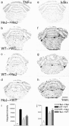
Early inflammatory response to LPS in CNS of chimeric mice. Film autoradiographs (a-h) show mRNA induction in brains of mice killed 2 h after intraperitoneal LPS. TNFα (a-d) and IκBα (e-h) mRNA in brainstem sections of the chimeric mice: HeJ → HeJ (a, e), WT → WT (b, f), WT → HeJ (c, g), and HeJ → WT (d, h). Induction of TNFα (i) and IκBα (j) mRNA in chimeric groups (details in key) were quantified by densitometry of film images. Data representing mean integrated density ± SEM for each mRNA were obtained from three or four mice per group. One-way ANOVA was followed by appropriate post hoc analysis to compare chimeric group means against the control WT → WT group. **p < 0.001.
Figure 6.
Figure 6.
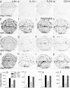
Late inflammatory response to LPS in CNS of chimeric mice. Representative film autoradiographs (a-p) show proinflammatory mRNA induction in brains of mice killed 6 h after intraperitoneal LPS. IκBα, IL-1β, TLR2 (i-l), and CD14 (m-p) mRNAs were detected by radioactive in situ hybridization on brainstem sections from chimeric mice: HeJ → HeJ (a, e, i, m), WT → WT (b, f, j, n), WT → HeJ (c, g, k, o), and HeJ → WT (d, h, l, p). Quantified expression levels are shown (q-t). IκBα (q), IL-1β (r), TLR2 (s), and CD14 (t) mRNA levels are plotted as mean integrated density ± SEM from three or four mice per chimeric class. Individual group means were compared against the control WT → WT group, and statistical significance was established by one-way ANOVA and appropriate post hoc tests. *p < 0.05; **p < 0.001.
Figure 7.
Figure 7.
Corticosterone (CORT) levels in serum from LPS-treated chimeric mice. Corticosterone was detected in serum from chimeric mice 2 or 6 h after an intraperitoneal injection of purified LPS (1 mg/kg), using a radioimmunoassay kit directed against murine corticosterone. Each bar represents mean ± SEM of values from three or four independent animals per group. Significant differences were established by two-way ANOVA, and post hoc tests were used to compare chimeric group means against the WT → WT control. **p < 0.001.

Comment in

Similar articles

Cited by

References

    1. Adams DH, Hanson GR, Keefe KA (2000) Cocaine and methamphetamine differentially affect opioid peptide mRNA expression in the striatum. J Neurochem 75: 2061-2070. - PubMed
    1. Berkenbosch F, van Oers J, del Rey A, Tilders F, Besedovsky H (1987) Corticotropin-releasing factor-producing neurons in the rat activated by interleukin-1. Science 238: 524-526. - PubMed
    1. Breder CD, Saper CB (1996) Expression of inducible cyclooxygenase mRNA in the mouse brain after systemic administration of bacterial lipopolysaccharide. Brain Res 713: 64-69. - PubMed
    1. Breder CD, Hazuka C, Ghayur T, Klug C, Huginin M, Yasuda K, Teng M, Saper CB (1994) Regional induction of tumor necrosis factor α expression in the mouse brain after systemic lipopolysaccharide administration. Proc Natl Acad Sci USA 91: 11393-11397. - PMC - PubMed
    1. Bsibsi M, Ravid R, Gveric D, van Noort JM (2002) Broad expression of Toll-like receptors in the human central nervous system. J Neuropathol Exp Neurol 61: 1013-1021. - PubMed

Publication types

MeSH terms