

A novel mechanism for desulfation of mucin: identification and cloning of a mucin-desulfating glycosidase (sulfoglycosidase) from Prevotella strain RS2
- PMID: 15716424
- PMCID: PMC1064001
- DOI: 10.1128/JB.187.5.1543-1551.2005
A novel mechanism for desulfation of mucin: identification and cloning of a mucin-desulfating glycosidase (sulfoglycosidase) from Prevotella strain RS2
Abstract
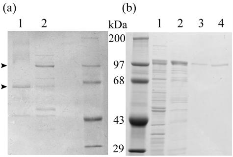
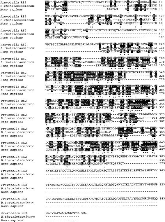
A novel enzyme which may be important in mucin degradation has been discovered in the mucin-utilizing anaerobe Prevotella strain RS2. This enzyme cleaves terminal 2-acetamido-2-deoxy-beta-D-glucopyranoside 6-sulfate (6-SO3-GlcNAc) residues from sulfomucin and from the model substrate 4-nitrophenyl 2-acetamido-2-deoxy-beta-D-glucopyranoside 6-sodium sulfate. The existence of this mucin-desulfating glycosidase (sulfoglycosidase) suggests an alternative mechanism by which this bacterium may desulfate sulfomucins, by glycosidic removal of a sulfated sugar from mucin oligosaccharide chains. Previously, mucin desulfation was thought to take place by the action of a specific desulfating enzyme, which then allowed glycosidases to remove desulfated sugar. Sulfate removal from sulfomucins is thought to be a rate-limiting step in mucin degradation by bacteria in the regions of the digestive tract with a significant bacterial flora. The sulfoglycosidase was induced by growth of the Prevotella strain on mucin and was purified 284-fold from periplasmic extracts. Tryptic digestion and sequencing of peptides from the 100-kDa protein enabled the sulfoglycosidase gene to be cloned and sequenced. Active recombinant enzyme was made in an Escherichia coli expression system. The sulfoglycosidase shows sequence similarity to hexosaminidases. The only other enzyme that has been shown to remove 6-SO3-GlcNAc from glycoside substrates is the human lysosomal enzyme beta-N-acetylhexosaminidase A, point mutations in which cause the inheritable, lysosomal storage disorder Tay-Sachs disease. The human enzyme removes GlcNAc from glycoside substrates also, in contrast to the Prevotella enzyme, which acts on a nonsulfated substrate at a rate that is only 1% of the rate observed with a sulfated substrate.
Figures
References
-
- Berkin, A., W. A. Szarek, and R. Kisilevsky. 2002. Synthesis and biological evaluation of a radiolabeled analog of methyl 2-acetamido-2,4-dideoxy-β-d-xylo-hexopyranoside directed towards influencing cellular glycosaminoglycan biosynthesis. Carbohydr. Res. 337:37-44. - PubMed
-
- Bernadi, G. 1971. Chromatography of proteins on hydroxyapatite. Methods Enzymol. 22:325-339. - PubMed
-
- Bradford, M. M. 1976. A rapid and sensitive method for the quantitation of microgram quantities of protein utilizing the principle of protein-dye binding. Anal. Biochem. 72:248-254. - PubMed
-
- Clinch, K., G. B. Evans, R. H. Furneaux, P. M. Rendle, P. L. Rhodes, A. M. Roberton, D. I. Rosendale, P. C. Tyler, and D. P. Wright. 2002. Synthesis and utility of sulfated chromogenic carbohydrate model substrates for measuring activities of mucin-desulfating enzymes. Carbohydr. Res. 337:1095-1111. - PubMed
Publication types
MeSH terms
Substances
Associated data
- Actions
LinkOut - more resources
Full Text Sources
Molecular Biology Databases
Research Materials
