. 2005 Apr;169(4):2061-74.
doi: 10.1534/genetics.104.036020. Epub 2005 Feb 16.

Gene expression analysis of the function of the male-specific lethal complex in Drosophila

Affiliations

Gene expression analysis of the function of the male-specific lethal complex in Drosophila

Manika Pal Bhadra et al. Genetics. 2005 Apr.

Abstract

Dosage compensation refers to the equal expression of X-linked genes despite the difference in copy number between the two sexes. The male-specific lethal (MSL) complex is concentrated on the X chromosome in males. A gene expression assay for embryos was developed to examine the function of this complex. In mutant male embryos without either the MSL complex or MOF histone acetylase, dosage compensation is retained but autosomal expression is increased. Dosage compensation is lost in the double-mutant embryos. In embryos in which the MSL complex and MOF are targeted to the X chromosomes in females, the results are consistent with previous surveys showing that in general the X expression remains unchanged, but autosomal expression is reduced. Mutations in the ISWI chromatin-remodeling component cause increases specifically of X-linked genes in males. Thus, the function of the MSL complex in conjunction with ISWI is postulated to override the effect on gene expression of high histone acetylation on the male X. The basic determinant of dosage compensation is suggested to be an inverse dosage effect produced by an imbalance of transcription factors on the X vs. the autosomes. The sequestration of the MSL complex to the male X may have evolved to counteract a similar effect on the autosomes and to prevent an overexpression of the X chromosome in males that would otherwise occur due to the high levels of histone acetylation.


Figures
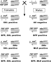
F<sc>igure</sc> 1.—
Figure 1.—
Genetic cross and resulting progeny to generate a segregating population for mle and mof. The cross +/Y; mle[pml8]/SM6a × y[1] mof[1]/FM7(GFP); mle[pml8]/mle[pml8] produces male progeny that are segregating for both mle and mof. The four types of males are normal [FM7(GFP)], mutant for mle alone (mle[pml8]/mle[pml8]), mutant for mof alone (mof[1]), and mutant for both mof and mle (mof[1]; mle[pml8]/mle[pml8]). The presence or absence of GFP, SXL, and MLE can be used to distinguish all four classes of males.
F<sc>igure</sc> 2.—
Figure 2.—
Development of a quantitative fluorescent gene expression assay. (A) Embryos stained with SXL antibodies and probed with antisense white and α-Gpdh RNAs. The anti-SXL labeling (blue) distinguishes the sex of the embryos. Females express SXL while males do not. The w mRNA hybridization was visualized using Cy3-conjugated UTP incorporated into the antisense probe. α-Gpdh mRNA was hybridized with an antisense probe labeled with Cy5-conjugated UTP. The pattern of Canton-S males and females is shown at the top. A segregating progeny of normal males and w67c23/+ heterozygous females, generated by crossing Canton-S females by males of a white deficiency strain (w67c23), are in the middle. A population of w−/+ female and w−/Y male embryos, which result from the reciprocal cross, are shown at the bottom. The genotype of each embryo is (a) Canton-S female, (b) Canton-S male, (c) w−/+ female, and (d) w− male. Bars, 50 μm. (B) A quantitative measurement of w, Adh, and α-Gpdh mRNA in embryos segregating for normal or null alleles of white or Adh, respectively. The white genotypes are described above. The heterozygous Adh normal/null genotype was produced by crossing Adhfn6 by Canton-S wild type, whereas the null homozygotes were obtained from an Adhfn6/ Adhfn6 stock. Normal males and females were obtained from the Canton-S stock. The Adh mRNA was detected with a digoxygenin-labeled antisense probe followed by incubation with Cy5-conjugated anti-dioxygenin antibodies. The same specific probe preparation for each of the three genes analyzed was used on all genotypes. Twenty-one embryos at approximately stage 13 were analyzed for each genotype. The amount of white, Adh, and α-Gpdh mRNA in each respective embryo was estimated by measuring the gray values per pixel using Metamorph software. The results indicate that the amount of mRNA detected by this technique is proportional to functional allele dosage and that a twofold difference in expression can be robustly distinguished. The results also illustrate that the quantitation of different genes (w vs. α-Gpdh) is independent of each other. (C) Comparison of null and antisense determinations of background. Normal embryos were probed for sense and antisense RNAs of white and Adh. In parallel, embryos homozygous for the respective null alleles were probed for mRNA. Embryos from three developmental stages are shown. The results illustrate that probing for antisense RNA is an effective background measurement. (D) Histograms of the comparison of background determinations via antisense probing or a null allele. The same digoxygenin-labeled probe preparation was used for analysis of normal and null genotypes for the respective genes. Twenty-one embryos were used to determine each mean. The results illustrate that the background determination by the two methods is comparable at the three developmental stages examined.
F<sc>igure</sc> 3.—
Figure 3.—
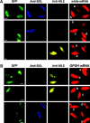
Gene expression analysis of embryos in the same microscopic field. (A) Effect of the mle and/or mof mutation on X-linked white transcripts in developing embryos. A collection of embryos segregating for the mle and mof mutations was probed with anti-SXL and anti-MLE antibodies followed by hybridization with an antisense white RNA probe. Each embryo was classified on the basis of GFP, SXL, and MLE. The genotypes are (a) mof/Y; mle/mle male, (b) GFP/+; mle/mle female, (c) y mof/+; mle/mle female, (d) GFP/Y; mle/mle male, and (e) y mof/Y; mle/+ male. Bars, 50 μm. (B) Effect of the mle and/or mof mutation on autosomal Gpdh RNA levels. Embryos were probed with two antibodies, SXL for sexing and MLE for genotyping, and also were hybridized with an antisense Gpdh RNA probe. The genotypes of each type of embryo are (a) GFP/Y; mle/mle male, (b) GFP/Y; mle/SM6a male, (c) mof/Y; mle/mle male, and (d) GFP/+; mle/mle female. Bars, 50 μm.
F<sc>igure</sc> 4.—
Figure 4.—
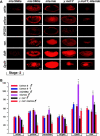
Embryo expression of the six selected genes analyzed. (A) A representative embryo at approximately stage 13 from each of the four male genotypes for each gene analyzed is shown. In addition, an early stage embryo is presented to illustrate the absence of maternal RNA. The sense mRNA determination represents the relative expression in each genotype. Probings for antisense RNA established the background hybridization. Bars, 50 μm. (B) Histogram of the gene expression in a population of embryos from Canton-S wild-type males and females as well as the mof/mle segregating population. Fifteen to 23 embryos were analyzed per genotype. Probing for antisense RNA served as the background for each gene, which was subtracted from the determination of each embryo. Those average values that differ from the normal male genotype at the 95% confidence level are marked by an asterisk.
F<sc>igure</sc> 5.—
Figure 5.—
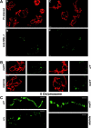
Distribution of the MSL complex in the mof mutant as revealed by MSL-1 labeling. (A) (a) A mixture of polytene chromosomes from mof males and mle/mle females stained with propidium iodide (PI). (b) The same nuclei probed for MSL-1. The females have no detectable MSL labeling on their chromosomes (Bhadra et al. 1999) and serve as a background control. The mof males show a destabilization of the MSL complex such that a low level of binding is now present on the autosomes, although a stronger labeling still occurs on the X. (c) A mixture of polytene chromosomes from mof and normal males stained with PI. (d) The same nuclei probed for MSL-1. The normal males show the usual strong labeling on the X chromosome and no detectable autosomal labeling. In contrast, the mof males exhibit some degree of MSL labeling on the autosomes. (B) Magnified views for comparison of single nuclei from the two mixtures in A. (C) Magnified views of the X chromosome from four genotypes. The normal male (+/Y) has strong labeling on the X. The normal female (+/+) (from a labeling not shown above) has a lower level of labeling, which is similar to the autosomes in the same nucleus (Hiebert and Birchler 1994). The mof male (mof/Y) retains strong binding at the high-affinity sites and weaker binding between them. The mle/mle female (mle/mle) shows no detectable labeling and illustrates that the signal between the strongly bound sites in the mof male is above background.
F<sc>igure</sc> 6.—
Figure 6.—
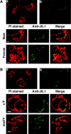
Distribution of JIL-1 kinase in the mof and normal males. (A) A mixture of polytene chromosomes from normal males and females was probed with antibody against JIL-1 kinase. (a) PI stained chromosomes. (b) Probings for JIL-1 kinase. Bottom, enlargement of a male and a female nucleus illustrating the enrichment of JIL-1 kinase on the X chromosome in males and the uniform distribution in females. (B) A mixture of polytene chromosomes from normal and mof mutant males stained with PI (a) and probed for JIL-1 kinase (b). Bottom, enlargements of single nuclei illustrating that the kinase is sequestered to the X in normal males, but released in the mof mutants to a uniform distribution on all chromosomes.
F<sc>igure</sc> 7.—
Figure 7.—
Gene expression analysis in Sxl embryos. From a cross of Sxlf1/FM7 GFP × Sxlfhv/Y, embryos were subjected to the fluorescent gene expression assay. This cross produces a segregating population in which mutant females are represented with greatly reduced SXL protein. This mutant combination was chosen to be in parallel with previous Northern analyses at the third instar larval stage (Bhadra et al. 2000). In the Sxl mutant cells, the MSL complex is formed and sequestered to the two X chromosomes, where the H4Lys16 acetylation is increased concomitant with a reduction of acetylation on the autosomes (Bhadra et al. 2000). Some genotypes in the progeny carry mutations for yellow and white, so they were not included in the analysis. A representative embryo (at approximately stage 13) for each gene in each genotype is shown. On the right, two fields of mixed stages probed for the X-linked 6Pgdh and the autosomal Rp49 genes are shown. a, FM7 GFP male; b, Sxlf1/Sxlfhv female; c, Sxlfhv/FM7 GFP female. A histogram of the seven genes analyzed is shown at the bottom right. A range of 10 to 36 embryos were analyzed for each value. The expression patterns of these representative genes are quite similar to a previous survey at the third instar larval stage (Bhadra et al. 2000). Generally, the X-linked genes are similar in expression to normal females, whereas autosomal gene expression is reduced. An asterisk denotes those means that are significantly different from the respective FM7-bearing genotype of the same sex.
F<sc>igure</sc> 8.—
Figure 8.—
Gene expression in the ISWI segregating population of embryos. Embryos were collected from a cross of w−/Y; CyO, GFP/ISWI1 × w−/w−; CyO GFP/ISWI2. Genotypes were classified on the basis of antibody labeling against SXL to determine sex and GFP excitation to determine the ISWI constitution. (A) Mixed stage embryos in the same field probed for the expression of the X-linked yellow gene. a, male ISWI mutant homozygote; b, female mutant homozygote; c, normal male. (B) Mixed stage embryos in the same field probed for the autosomal gene, α-Gpdh. a, male mutant homozygote; b, normal female; c, normal male. (C) Representative embryos at approximately stage 13 for four X-linked genes (y, G6pdh, 6Pgdh, and v) and two autosomal genes (Adh and α-Gpdh). The white gene expression was not tested because the X chromosome in these stocks carries a mutant allele. (D) Histogram of the mean gray values of embryos at approximately stage 13 from the segregating ISWI population. A range of 10 to 20 embryos were analyzed for each mean. An asterisk denotes those means that are significantly different from the normal genotype of the same sex.
F<sc>igure</sc> 9.—
Figure 9.—
Summary of gene expression. The evolutionary progenitor situation to the heteromorphic sex chromosomes in Drosophila is assumed to involve a pair of homologous chromosomes with similar gene expression between the sexes. The current karyotype shows dosage compensation of the single X chromosome in males compared to two in females and nearly equal autosomal expression. Because the gene expression in the msl mutants shows hyperactivation of both the X and the autosomes, it is postulated that a global inverse dosage effect, typical of monosomic situations, is operating and that the MSL sequestration evolved to counteract this dosage effect on the autosomes. Retention of dosage compensation in the mof mutant males, but not in the double mutant, indicates that the MSL complex overrides the impact of histone acetylation, in this case when acetylation on the X is reduced. The loss of dosage compensation in the double-mutant mof; mle indicates that the MSL chromatin remodeling complex is necessary for the inverse dosage effect to operate. The failure to increase X expression when the MSL complex and increased H4 acetylation is targeted to the X's in females indicates the override also operates with increased acetylation. The overcompensation of the male X in the ISWI mutants suggests that this chromatin remodeling subunit is required for the override process.

Similar articles

Cited by

References

    1. Akhtar, A., and P. B. Becker, 2000. Activation of transcription through histone H4 acetylation by MOF, an acetyltransferase essential for dosage compensation in Drosophila. Mol. Cell 5: 367–375. - PubMed
    1. Arkhipova, I., J. Li and M. Meselson, 1997. On the mode of gene-dosage compensation in Drosophila. Genetics 145: 729–736. - PMC - PubMed
    1. Badenhorst, P., M. Voas, I. Rebay and C. Wu, 2002. Biological functions of the ISWI chromatin remodeling complex NURF. Genes Dev. 16: 3186–3198. - PMC - PubMed
    1. Bahn, S., M. Mimmack, M. Ryan, M. A. Caldwell, E. Jauniaux et al., 2002. Neuronal target genes of the neuron-restrictive silencer factor in neurospheres derived from fetuses with Down's syndrome: a gene expression study. Lancet 359: 310–315. - PubMed
    1. Belote, J. M., and J. C. Lucchesi, 1980. Control of X chromosome transcription by the maleless gene in Drosophila. Nature 285: 573–575. - PubMed

Publication types

MeSH terms

LinkOut - more resources