Review
. 2004 Jan;1(1):17-25.
doi: 10.1602/neurorx.1.1.17.

Neuronal death/survival signaling pathways in cerebral ischemia

Affiliations
Review

Neuronal death/survival signaling pathways in cerebral ischemia

Taku Sugawara et al. NeuroRx. 2004 Jan.

Abstract

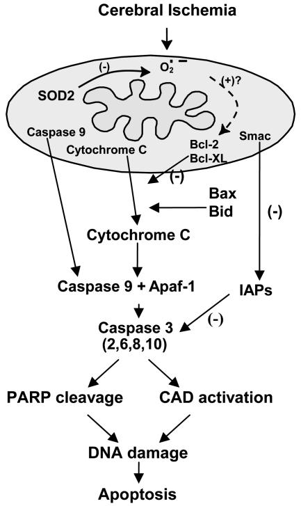
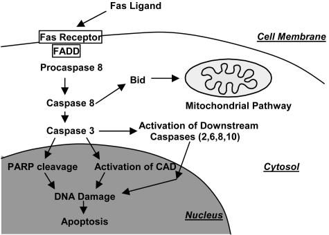
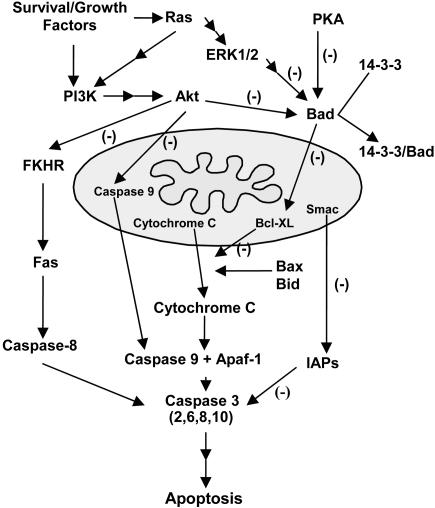
Cumulative evidence suggests that apoptosis plays a pivotal role in cell death in vitro after hypoxia. Apoptotic cell death pathways have also been implicated in ischemic cerebral injury in in vivo ischemia models. Experimental ischemia and reperfusion models, such as transient focal/global ischemia in rodents, have been thoroughly studied and the numerous reports suggest the involvement of cell survival/death signaling pathways in the pathogenesis of apoptotic cell death in ischemic lesions. In these models, reoxygenation during reperfusion provides a substrate for numerous enzymatic oxidation reactions. Oxygen radicals damage cellular lipids, proteins and nucleic acids, and initiate cell signaling pathways after cerebral ischemia. Genetic manipulation of intrinsic antioxidants and factors in the signaling pathways has provided substantial understanding of the mechanisms involved in cell death/survival signaling pathways and the role of oxygen radicals in ischemic cerebral injury. Future studies of these pathways may provide novel therapeutic strategies in clinical stroke.


Figures

FIG. 1.
FIG. 1.
Mitochondria as targets for oxidative stress signaling after cerebral ischemia. Cerebral ischemia and reperfusion generate ROS within mitochondria, which then signal the release of cytochrome c by mechanisms that may be related to the Bcl-2 family proteins, Bcl-2, Bcl-XL, Bax, and Bid. Cytochrome c, once released, binds to Apaf-1 followed by caspase-9 to form a complex that subsequently activates caspase-3 and other caspases, such as caspase-2, -6, -8, and -10. The IAP family suppresses apoptosis by preventing the activation of procaspases and also inhibits the enzymatic activity of active caspases; Smac is also released by apoptotic stimuli and binds IAPs, thereby promoting activation of caspase-3. Activated caspase-3 is known to cleave many nuclear DNA repair enzymes, such as PARP and to activate CAD, which then leads to nuclear DNA damage without repair, resulting in apoptosis.
FIG. 2.
FIG. 2.
Fas receptor pathway of apoptosis. The extracellular Fas ligand first binds to a receptor, and then an adaptor molecule, FADD protein, activates procaspase-8. Then, caspase-8 activates caspase-3 and this effector caspase cleaves PARP and activates CAD, leading to DNA damage and cell death. In the middle of this pathway, caspase-3 uses downstream caspases as in the mitochondrial pathway. Caspase-8 is also able to truncate and activate one of the Bcl-2 family proteins, Bid, and initiates the mitochondrial pathway of apoptosis.
FIG. 3.
FIG. 3.
Cell survival signaling pathways. Three major pathways that inhibit Bad are shown. Bad is an important proapoptotic member of the Bcl-2 family that links cell survival and apoptosis pathways. Bad promotes the release of cytochrome c by inhibiting the antiapoptotic effects of Bcl-XL. The phosphatidylinositol-3 kinase (PI3K) pathway is activated by survival/growth factors and leads to activation of Akt. Akt inhibits the Forkhead family of transcription factors (FKHR), caspase-9, and Bad, and ultimately leads to inhibition of both mitochondrial and Fas receptor pathways of apoptosis. ERK1/2 activation through active Ras and PKA also inhibit Bad and thereby block cytochrome c release by Bcl-XL inhibition.

References

    1. Chan PH. Reactive oxygen radicals in signaling and damage in the ischemic brain. J Cereb Blood Flow Metab 21: 2–14, 2001. - PubMed
    1. Chan PH. Oxygen radicals in focal cerebral ischemia. Brain Pathol 4: 59–65, 1994. - PubMed
    1. Lewen A, Matz P, Chan PH. Free radical pathways in CNS injury. J Neurotrauma 17: 871–890, 2000. - PubMed
    1. Sugawara T, Lewen A, Gasche Y, Yu F, Chan PH. Overexpression of SOD1 protects vulnerable motor neurons after spinal cord injury by attenuating mitochondrial cytochrome c release. FASEB J 16: 1997–1999, 2002. - PubMed
    1. Liu R, Althaus JS, Ellerbrock BR, Becker DA, Gurney ME. Enhanced oxygen radical production in a transgenic mouse model of familial amyotrophic lateral sclerosis. Ann Neurol 44: 763–770, 1998. - PubMed

Publication types

Substances