Review
. 2004 Oct;1(4):424-51.
doi: 10.1602/neurorx.1.4.424.

Cellular transplantation strategies for spinal cord injury and translational neurobiology

Affiliations
Review

Cellular transplantation strategies for spinal cord injury and translational neurobiology

Paul J Reier. NeuroRx. 2004 Oct.

Abstract

Basic science advances in spinal cord injury and regeneration research have led to a variety of novel experimental therapeutics designed to promote functionally effective axonal regrowth and sprouting. Among these interventions are cell-based approaches involving transplantation of neural and non-neural tissue elements that have potential for restoring damaged neural pathways or reconstructing intraspinal synaptic circuitries by either regeneration or neuronal/glial replacement. Notably, some of these strategies (e.g., grafts of peripheral nerve tissue, olfactory ensheathing glia, activated macrophages, marrow stromal cells, myelin-forming oligodendrocyte precursors or stem cells, and fetal spinal cord tissue) have already been translated to the clinical arena, whereas others have imminent likelihood of bench-to-bedside application. Although this progress has generated considerable enthusiasm about treating what once was thought to be a totally incurable condition, there are many issues to be considered relative to treatment safety and efficacy. The following review reflects on different experimental applications of intraspinal transplantation with consideration of the underlying pathological, pathophysiological, functional, and neuroplastic responses to spinal trauma that such treatments may target along with related issues of procedural and biological safety. The discussion then moves to an overview of ongoing and completed clinical trials to date. The pros and cons of these endeavors are considered, as well as what has been learned from them. Attention is primarily directed at preclinical animal modeling and the importance of patterning clinical trials, as much as possible, according to laboratory experiences.


Figures
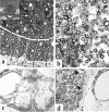
FIG. 1.
FIG. 1.
a-d: Partial preservation of myelinated fibers is illustrated in plastic semithin sections and a MR transverse slice obtained from a human subject (a, inset). a: A region of ventrolateral white matter is shown at a contusion epicenter site 3 weeks postinjury in an adult rat spinal cord. This region is subdivided (dashed white line) into a more centrally located zone that is undergoing extensive fiber degeneration and a more peripheral region (pial surface indicated by arrows), in which a rim of some spared fibers is seen. A more severe injury is illustrated in c. In this transverse section of the lesion epicenter of an adult cat spinal cord, as seen 4 months after a static load compression injury, nearly all of the spinal cord has deteriorated except for regions in which a few axons are present subjacent to the surrounding pia. The boxed area represents one such fiber zone that is shown at higher magnification in d. In regions of spared white matter (b and d), there can be extensive demyelination and remyelination noted (arrows point to some examples). These regions also may appear less compacted because of associated fiber loss and edema. The MR image (inset) shows a human correlate of what is illustrated in histological sections. A rim of white matter can be identified surrounding a large central cyst at T5. Preop, preoperative.
FIG. 2.
FIG. 2.
a: A plastic semithin section, showing in transverse plane a static-load compression epicenter site in an adult cat spinal cord. A central region of highly partitioned cystic cavitation is indicated by arrows. The shell of surrounding “preserved” white matter has features similar to those shown in Figure 1, a and b. b: A “living” correlate of this image is seen in this syringoscopic view of a highly compartmentalized cyst (arrows) in a patient during an exploratory phase of intraspinal surgery (courtesy of Drs. Edward Wirth III and Richard Fessler).
FIG. 3.
FIG. 3.
Motoneuron loss after a spinal injury may not exhibit profound clinical symptoms except under more demanding conditions. This figure illustrates phrenic motoneuron pool (i.e., C3-C5) utilization in adult rats as exemplified by diaphragm EMG amplitudes during augmented breaths (i.e., equivalent to a sigh). Under normal breathing conditions, only 50% of the phrenic motoneuron pool is used (dashed line); however, when augmented breathing is induced, rats will recruit an additional 10-20% of available phrenic motoneurons. After a C4-C5 contusion injury, respiratory function is not overtly different from that seen in controls under normal conditions. However, when faced with respiratory challenge, those animals recruit virtually all of the remaining phrenic motoneuron pool (data obtained in collaboration with Dr. Donald Bolser). Thus, from a human perspective, even when a cervically injured individual may not require ventilatory assistance, respiratory performance is severely compromised, thus leading to respiratory weakness evident, for example, in speech or cough.
FIG. 4.
FIG. 4.
Extensive white matter damage is seen in these micrographs taken from the epicenter of a static load compression injury to an adult cat spinal cord. Months after that injury, the white matter is replaced by an amorphic cellular terrain in which large fascicular structures are seen (a). At a higher magnification (b), these structures are bundles of axons that are myelinated by Schwann cells. Some individual fibers are seen in the surrounding tissue parenchyma. As discussed in the accompanying text, Schwann cell infiltration of the injured spinal cord is a common feature in animals and humans and represents one form of potential self-repair that can be augmented by grafts of Schwann cells and other cell types (e.g., olfactory ensheathing glia).
FIG. 5.
FIG. 5.
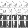
Gait records obtained from one subject enrolled in the Gainesville human fetal spinal cord trial described in the accompanying text. Overground locomotion was recorded at normal pace (0.14 m/sec) and fast pace (0.19 m/sec) before surgery and then 3 months post-transplantation (normal pace, 0.55 m/sec; fast pace, 0.75 m/sec). In addition to achieving faster locomotion after surgery, this individual also showed improved performance relative to a preoperative foot-drop (arrows) which were noticeably absent after transplantation (data obtained by Dr. Andrea Behrman). Preop, preoperative; Postop, postoperative.
FIG. 6.
FIG. 6.
Translational correspondence between preclinical locomotor performance in a cat before (row A) and after (row B) intraspinal fetal transplant surgery compared with a human subject before (row C) and after (row D) human fetal cell grafting. Before transplantation, both the cat and human subject show a pronounced foot drop (see FIG. 5 for the person in rows C and D) with improved locomotor performance after surgery.
FIG. 7.
FIG. 7.
A transverse section of an adult rat spinal cord in which a transplant was made on one side using human fetal spinal cord (huFSC) tissue remaining after one of the grafting sessions in the Gainesville post-traumatic syringomyelia/fetal cell transplantation trial. Host gray matter (GMh) and central canal (Cc) are identified for reference. This section was obtained 1 month after transplantation. Human fetal tissue xenografts mature slowly, and thus the transplant illustrated is primarily comprised of germinal neuroepithelial cells and a few large blood vessels. This approach provided evidence of the viability of donor tissue used on each patient enrolled in the specified trial.
FIG. 8.
FIG. 8.
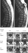
Sagittal (a) and transverse (b) MR images from the subject whose locomotor function is illustrated in Figures 5 and 6. This person’s injury occurred 30 years before the transplantation procedure. Her presenting neurological symptoms before being enrolled were associated with an expanding syrinx at the Th5-Th6 interface [preoperative (Preop) image] where a pronounced kyphosis also was evident. One year after surgery, much of the region of cavitation was obliterated, although the host-graft interfaces were not obvious. Comparison with images at 3 months after grafting suggested some cavitation being seen in the region of grafting that was not apparent earlier, although no declining neurological symptoms were detected. Transverse images revealed substantial host tissue preservation before grafting, which represented a substantial risk. Images obtained 6 weeks and 12 months after transplantation suggested significant filling of the cyst; however, changes in graft appearance could be seen between these two times in some sections (e.g., at Th5-Th6).
FIG. 9.
FIG. 9.
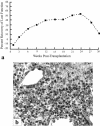
In a, the post-transplantation functional recovery pattern of adult cats with chronic compression lesions of the spinal cord and grafts of fetal cat spinal cord tissue is shown. Data are based on scoring that established an index of locomotor function. Evaluations were made before and after transplantation, and the postgrafting data are presented as a percentage change relative to pregraft baseline (data extracted from Anderson et al.320). Five animals received transplants. One animal per postgraft interval was killed at 9, 18, and 24 weeks after grafting. Two animals survived until 30 weeks. One of these cats had a graft undergoing rejection (b) and showed a significant behavioral decline, which contributed to the progressive functional regression shown in this graph between 24 and 30 weeks. Immunological deletion thus suggested some graft-mediated functional recovery, especially because no overt pathology was seen in neighboring host tissue. As noted in the text, a similar pattern is now being seen in one huFSC graft recipient.

References

    1. Kao CC, Bunge RP, Reier PJ. Spinal cord reconstruction. New York: Raven Press, 1983.
    1. Sugar O, Gerard RW. Spinal cord regeneration in the rat. J Neurophysiol 3: 1–19, 1940.
    1. Hulsebosch CE. Recent advances in pathophysiology and treatment of spinal cord injury. Adv Physiol Educ 26: 238–255, 2002. - PubMed
    1. DeFelipe J, Jones EG. Cajal’s degeneration and regeneration of the nervous system (May RM, translator). New York: Oxford University Press, 1991.
    1. David S, Aguayo AJ. Axonal elongation into peripheral nervous system “bridges” after central nervous system injury in adult rats. Science 214: 931–933, 1981. - PubMed

Publication types