Review
. 2005 Jan;2(1):129-38.
doi: 10.1602/neurorx.2.1.129.

Tyrosine hydroxylase replacement in experimental Parkinson's disease with transvascular gene therapy

Affiliations
Review

Tyrosine hydroxylase replacement in experimental Parkinson's disease with transvascular gene therapy

William M Pardridge. NeuroRx. 2005 Jan.

Abstract

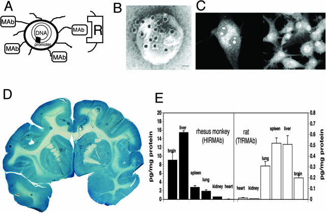
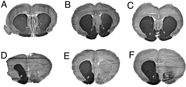
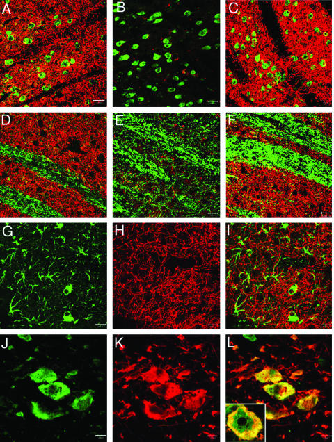
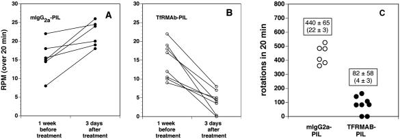
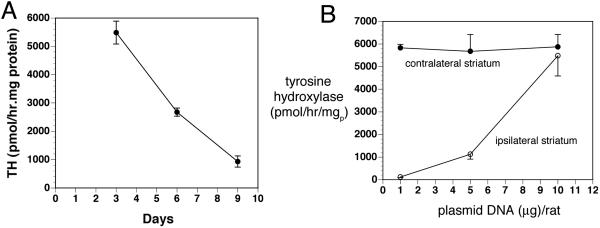
Transvascular gene therapy of Parkinson's disease (PD) is a new approach to the gene therapy of PD and involves the global distribution of a therapeutic gene to brain after an intravenous administration and transport across the blood-brain barrier (BBB). This is enabled with the development of a nonviral gene transfer technology that encapsulates plasmid DNA inside pegylated immunoliposomes or PILs. An 85- to 100-nm liposome carries the DNA inside the nanocontainer, and the liposome surface is conjugated with several thousand strands of 2000-Da polyethyleneglycol (PEG). This PEGylation of the liposome stabilizes the structure in the blood stream. The liposome is targeted across the BBB via attachment to the tips of 1-2% of the PEG strands of a receptor-specific monoclonal antibody (mAb) directed at a BBB receptor, such as the insulin receptor or transferrin receptor (TfR). Owing to the expression of the insulin receptor or the TfR on both the BBB and the neuronal plasma membrane, the PIL is able to reach the neuronal nuclear compartment from the circulation. Brain-specific expression is possible with the combined use of the PIL gene transfer technology and brain-specific gene promoters. In the 6-hydroxydopamine rat model of experimental PD, striatal tyrosine hydroxylase (TH) activity is completely normalized after an intravenous administration of TfRmAb-targeted PILs carrying a TH expression plasmid. A treatment for PD may be possible with dual gene therapy that seeks both to replace striatal TH gene expression with TH gene therapy, and to halt or reverse neurodegeneration of the nigro-striatal tract with neurotrophin gene therapy.


Figures

FIG. 1.
FIG. 1.
A: Diagram of a super-coiled expression plasmid DNA encapsulated in an 85 nm pegylated PIL targeted to a cell membrane receptor (R) with a receptor-specific, endocytosing mAb. Tissue-specific expression of the plasmid can be controlled by the promoter inserted 5′ of the gene. Panels A and B are reproduced with permission from Zhang et al. Intravenous nonviral gene therapy causes normalization of striatal tyrosine hydroxylase and reversal of motor impairment in experimental parkinsonism. Hum Gene Ther 14:1–12. Copyright © 2003, Mary Ann Leibert, Inc. All rights reserved. B: Transmission electron microscopy of a PIL. The mAb molecule tethered to the tips of the 2000-Da PEG is bound by a conjugate of 10 nm gold and a secondary antibody. The position of the gold particles shows the relationship of the PEG extended mAb and the liposome. Magnification bar = 20 nm. C: Confocal microscopy of U87 human glioma cells after either a 3-h (left panel) or a 24-h (right panel) incubation of fluorescein conjugated clone 882 DNA (fluoro-DNA) encapsulated within HIRmAb-PILs. The inverted grayscale image is shown. There is primarily cytoplasmic accumulation of the fluoro-DNA at 3 h, whereas the fluoro-DNA is largely confined to the nuclear compartment at 24 h. Fluoro-DNA entrapped within intranuclear vesicles is visible at both 3 and 24 h. Panel C is reproduced with permission from Zhang et al. Receptor-mediated delivery of an antisense gene to human brain cancer cells. J Gene Med 4:183–194. Copyright © 2002, John Wiley & Sons, Ltd. All rights reserved. D: β-Galactosidase histochemistry of brain removed 48 h after the intravenous injection of a β-galactosidase expression plasmid encapsulated in HIRmAb-PILs in the adult rhesus monkey. Panels D and E are reproduced with permission from Zhang et al. Global non-viral gene transfer to the primate brain following intravenous administration. Mol Ther 7:11–18. Copyright © 2003, Academic Press. All rights reserved. E: Luciferase gene expression in the brain and other organs of the adult rhesus monkey (left panel) and adult rat (right panel) measured at 48 h after a single intravenous injection of the PIL carrying the plasmid DNA. Data are mean ± SEM. The plasmid DNA encoding the luciferase gene used in either species is clone 790, which is driven by the SV40 promoter. The PIL carrying the DNA was targeted to primate organs with an HIRmAb and to rat organs with a TfRmAb.
FIG. 2.
FIG. 2.
Tyrosine hydroxylase immunocytochemistry of rat brain removed 72 h after a single intravenous injection of 10 μg per rat of clone 951 plasmid DNA encapsulated in PIL targeted with either the TfRmAb (panels A–C) or with the mouse IgG2a isotype control (panels D–F). Coronal sections are shown for three different rats from each of the two treatment groups. Clone 951 is the TH expression plasmid under the influence of the human GFAP promoter. The 6-hydroxydopamine was injected in the medial forebrain bundle of the right hemisphere, which corresponds to right side of the figure. Sections are not counterstained. Reproduced with permission from Zhang et al. Normalization of striatal tyrosine hydroxylase and reversal of motor impairment in experimental parkonsinism with intravenous nonviral gene therapy and a brain-specific promotor. Hum Gene Ther 15:339–350. Copyright © 2004, Mary Ann Liebert, Inc. All rights reserved.
FIG. 3.
FIG. 3.
Confocal microscopy of striatum and substantia nigra in 6-hydroxydopamine-lesioned rats sacrificed at 3 days after intravenous injection of clone 951 plasmid DNA encapsulated in PILs targeted either with mouse IgG2a (panels B and E) or with the TfRmAb (panels A, C, D, F, and G–L). Panels A and D are from the striatum contralateral to the lesion, and panels B, C, E, F, and G–I are from the striatum ipsilateral to toxin injection. Panels J to L are from the substantia nigra ipsilateral to the lesion. Panels A–C show striatum colabeled with a mouse monoclonal antibody to NeuN (green) and a rabbit polyclonal antibody to TH (red). Panels D–F show striatum colabeled with a mouse monoclonal antibody to the 200-kDa neurofilament protein (green) and a rabbit polyclonal antibody to TH (red). The magnification bar in panel A is 20 μm. All images are three-dimensional projection views of multiple planar images. The yellow color is an artifact from the three-dimensional projection because there was no overlap observed in the single planar views. Panels G and J show immune staining (green channel) with monoclonal antibodies to GFAP and NeuN, respectively. Panels H and K show immune staining (red channel) with a rabbit polyclonal antibody to TH. The overlap image of TH and GFAP in striatum is shown in panel I; the overlap image of TH and NeuN in substantia nigra is shown in panel L. The inset of panel L is a 100× oil immersion view of colabeling of TH (red), NeuN (green), and the overlap (yellow) in a neuron in the substantia nigra. The magnification bars in panels G and J are 20 and 10 μm, respectively. All images are three-dimensional projection views of multiple planar images. Reproduced with permission from Zhang et al. Normalization of striatal tyrosine hydroxylase and reversal of motor impairment in experimental parkinsonism with intravenous nonviral gene therapy and a brain-specific promotor. Hum Gene Ther 15:339–350. Copyright © 2004, Mary Ann Liebert, Inc. All rights reserved.
FIG. 4.
FIG. 4.
A: Apomorphine-induced rotations per minute (RPM) over a 20-min period measured in individual rats at 1 week before treatment and at 3 days after a single intravenous injection of 10 μg per rat of clone 951 plasmid DNA encapsulated in a PIL targeted with the mouse IgG2a isotype control antibody. B: Apomorphine-induced RPM over a 20 min period measured in individual rats at 1 week before treatment and at 3 days after a single intravenous injection of 10 μg per rat of clone 951 plasmid DNA encapsulated in a PIL targeted with the TfRmAb. C: Comparison of the total rotations in the two groups at 3 days after treatment. The average RPM is 22 ± 3 and 4 ± 3 (mean ± SD) in animals treated with the mIgG2a-PIL and the TfRmAb-PIL, respectively. The difference in rotation between the two groups is significant at the p < 0.005 level. Reproduced with permission from Zhang et al. Normalization of striatal tyrosine hydroxylase and reversal of motor impairment in experimental parkonsinism with intravenous nonviral gene therapy and a brain-specific promotor. Hum Gene Ther 15:339–350. Copyright © 2004, Mary Ann Liebert, Inc. All rights reserved.
FIG. 5.
FIG. 5.
A: The striatal TH activity ipsilateral to the 6-hydroxydopamine lesion is plotted versus time after a single intravenous injection of 10 μg/rat of clone 877 plasmid DNA encapsulated in the TfRmAb-PIL at day 0. Data are mean ± SD (n = 3 rats per point). Clone 877 is the TH expression plasmid under the influence of the SV40 promoter. B: The striatal TH activity either ipsilateral or contralateral to the 6-hydroxydopamine lesion is plotted versus the dose of clone 877 plasmid DNA encapsulated in the TfRmAb-PIL. Data are mean ± SD (n = 3 rats per point). Striatal TH was measured at 3 days after the single intravenous administration of the DNA. Reproduced with permission from Zhang et al. Intravenous nonviral gene therapy causes normalization of striatal tyrosine hydroxylase and reversal of motor impairment in experimental parkinsonism. Hum Gene Ther 14:1–12. Copyright © 2003, Mary Ann Liebert, Inc. All rights reserved.

Similar articles

Cited by

References

    1. Shastry BS. Parkinson disease: etiology, pathogenesis and future of gene therapy. Neurosci Res 41: 5–12, 2001. - PubMed
    1. Booij J, Bergmans P, Winogrodzka A, Speelman JD, Wolters EC. Imaging of dopamine transporters with [123I]FP-CIT SPECT does not suggest a significant effect of age on the symptomatic threshold of disease in Parkinson's disease. Synapse 39: 101–108, 2001. - PubMed
    1. Boado RJ, Li JY, Nagaya M, Zhang C, Pardridge WM. Selective expression of the large neutral amino acid transporter at the blood-brain barrier. Proc Natl Acad Sci USA 96: 12079–12084, 1999. - PMC - PubMed
    1. Kordower JH, Emborg ME, Bloch J, Ma SY, Chu Y, Leventhal L, et al. Neurodegeneration prevented by lentiviral vector delivery of GDNF in primate models of Parkinson's disease. Science 290: 767–773, 2000. - PubMed
    1. Dewey RA, Morrissey G, Cowsill CM, Stone D, Bolognani F, Dodd NJ, et al. Chronic brain inflammation and persistent herpes simplex virus 1 thymidine kinase expression in survivors of syngeneic glioma treated by adenovirus-mediated gene therapy: implications for clinical trials. Nat Med 5: 1256–1263, 1999. - PubMed

Publication types

MeSH terms