

Identification, characterization and structure of a new Delta class glutathione transferase isoenzyme
- PMID: 15717864
- PMCID: PMC1183455
- DOI: 10.1042/BJ20042015
Identification, characterization and structure of a new Delta class glutathione transferase isoenzyme
Abstract
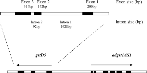
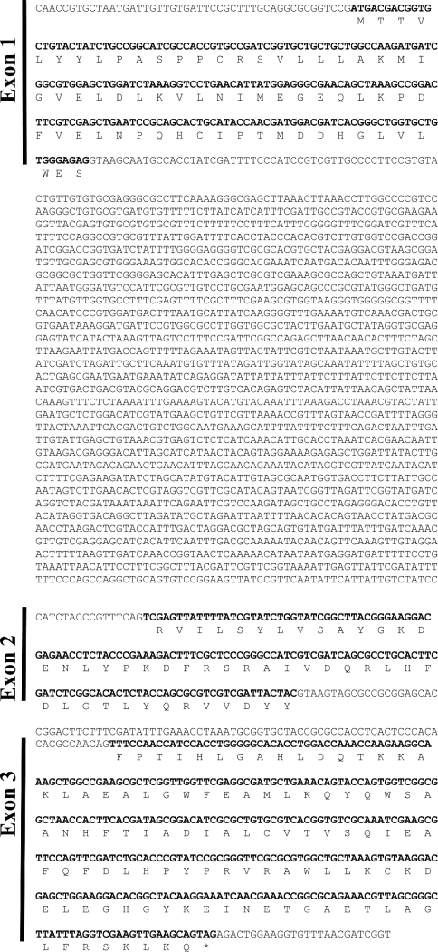
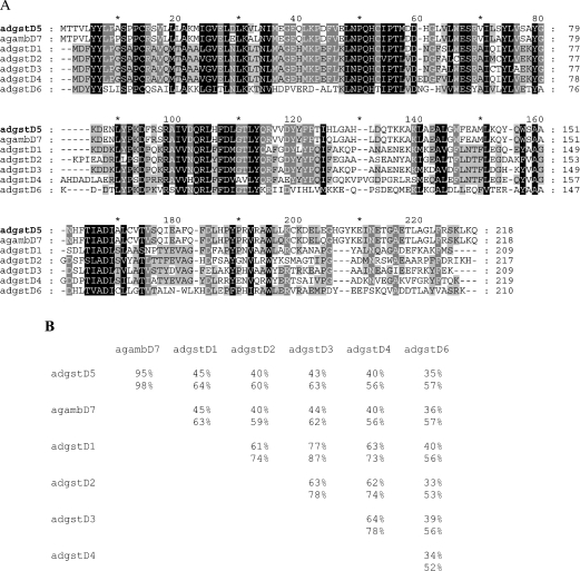
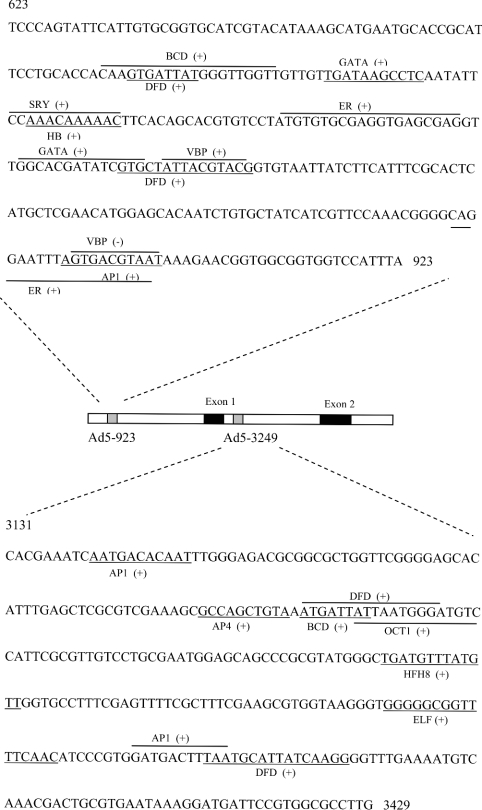
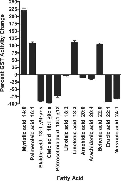
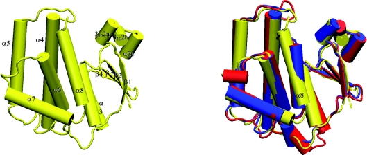
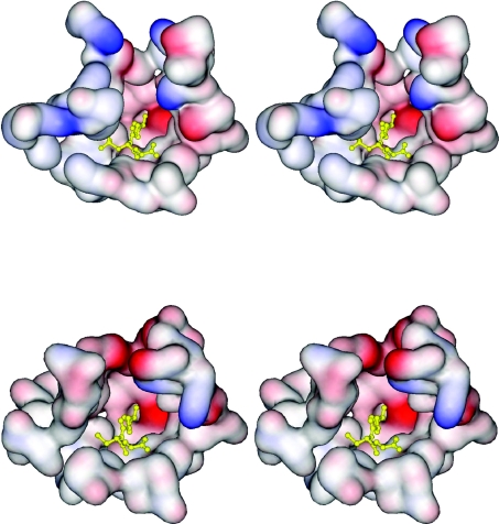
The insect GST (glutathione transferase) supergene family encodes a varied group of proteins belonging to at least six individual classes. Interest in insect GSTs has focused on their role in conferring insecticide resistance. Previously from the mosquito malaria vector Anopheles dirus, two genes encoding five Delta class GSTs have been characterized for structural as well as enzyme activities. We have obtained a new Delta class GST gene and isoenzyme from A. dirus, which we name adGSTD5-5. The adGSTD5-5 isoenzyme was identified and was only detectably expressed in A. dirus adult females. A putative promoter analysis suggests that this GST has an involvement in oogenesis. The enzyme displayed little activity for classical GST substrates, although it possessed the greatest activity for DDT [1,1,1-trichloro-2,2-bis-(p-chlorophenyl)ethane] observed for Delta GSTs. However, GST activity was inhibited or enhanced in the presence of various fatty acids, suggesting that the enzyme may be modulated by fatty acids. We obtained a crystal structure for adGSTD5-5 and compared it with other Delta GSTs, which showed that adGSTD5-5 possesses an elongated and more polar active-site topology.
Figures

Similar articles
-
Cloning, expression and characterization of an insect class I glutathione S-transferase from Anopheles dirus species B.Insect Biochem Mol Biol. 1998 May-Jun;28(5-6):321-9. doi: 10.1016/s0965-1748(98)00006-x. Insect Biochem Mol Biol. 1998. PMID: 9692235
-
Structure of an insect delta-class glutathione S-transferase from a DDT-resistant strain of the malaria vector Anopheles gambiae.Acta Crystallogr D Biol Crystallogr. 2003 Dec;59(Pt 12):2211-7. doi: 10.1107/s0907444903018493. Epub 2003 Nov 27. Acta Crystallogr D Biol Crystallogr. 2003. PMID: 14646079
-
Identification of a novel class of insect glutathione S-transferases involved in resistance to DDT in the malaria vector Anopheles gambiae.Biochem J. 2001 Oct 15;359(Pt 2):295-304. doi: 10.1042/0264-6021:3590295. Biochem J. 2001. PMID: 11583575 Free PMC article.
-
Mosquito glutathione transferases.Methods Enzymol. 2005;401:226-41. doi: 10.1016/S0076-6879(05)01014-1. Methods Enzymol. 2005. PMID: 16399389 Review.
-
Characterization of the omega class of glutathione transferases.Methods Enzymol. 2005;401:78-99. doi: 10.1016/S0076-6879(05)01005-0. Methods Enzymol. 2005. PMID: 16399380 Review.
Cited by
-
Association of glutathione S-transferase T1, M1, and P1 polymorphisms in the breast cancer risk: a meta-analysis.Ther Clin Risk Manag. 2016 May 12;12:763-9. doi: 10.2147/TCRM.S104339. eCollection 2016. Ther Clin Risk Manag. 2016. PMID: 27274261 Free PMC article.
-
Characterization and functional analysis of four glutathione S-transferases from the migratory locust, Locusta migratoria.PLoS One. 2013;8(3):e58410. doi: 10.1371/journal.pone.0058410. Epub 2013 Mar 7. PLoS One. 2013. PMID: 23505503 Free PMC article.
-
Expression Patterns of Drosophila Melanogaster Glutathione Transferases.Insects. 2022 Jul 7;13(7):612. doi: 10.3390/insects13070612. Insects. 2022. PMID: 35886788 Free PMC article.
-
Tetrahymena thermophila glutathione-S-transferase superfamily: an eco-paralogs gene network differentially responding to various environmental abiotic stressors and an update on this gene family in ciliates.Front Genet. 2025 Mar 7;16:1538168. doi: 10.3389/fgene.2025.1538168. eCollection 2025. Front Genet. 2025. PMID: 40125531 Free PMC article.
-
Comparison of epsilon- and delta-class glutathione S-transferases: the crystal structures of the glutathione S-transferases DmGSTE6 and DmGSTE7 from Drosophila melanogaster.Acta Crystallogr D Biol Crystallogr. 2015 Oct;71(Pt 10):2089-98. doi: 10.1107/S1399004715013929. Epub 2015 Sep 26. Acta Crystallogr D Biol Crystallogr. 2015. PMID: 26457432 Free PMC article.
References
-
- Prapanthadara L., Hemingway J., Ketterman A. J. Partial purification and characterization of glutathione S-transferases involved in DDT resistance from the mosquito Anopheles gambiae. Pestic. Biochem. Physiol. 1993;47:119–133.
Publication types
MeSH terms
Substances
Associated data
- Actions
LinkOut - more resources
Full Text Sources
Other Literature Sources
Research Materials
Miscellaneous