. 2005 Mar 9;24(5):1068-78.
doi: 10.1038/sj.emboj.7600589. Epub 2005 Feb 17.

Bcl-2 enhances Ca(2+) signaling to support the intrinsic regenerative capacity of CNS axons

Affiliations

Bcl-2 enhances Ca(2+) signaling to support the intrinsic regenerative capacity of CNS axons

Jianwei Jiao et al. EMBO J. .

Abstract

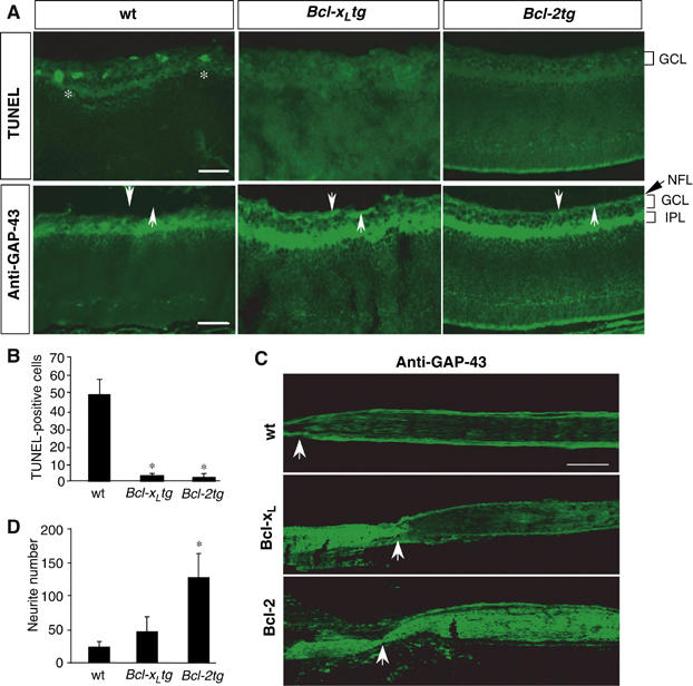
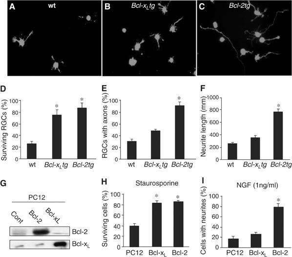
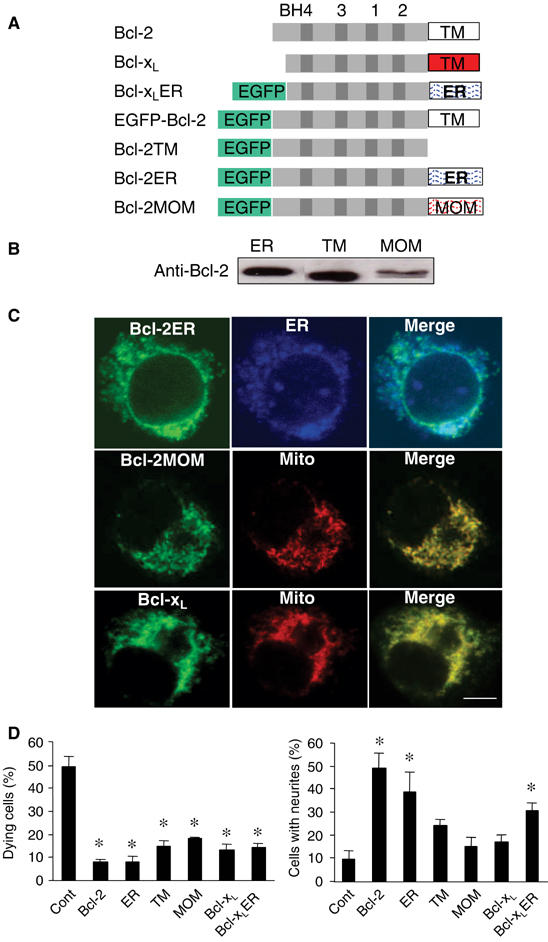
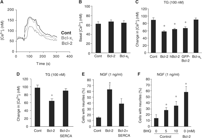
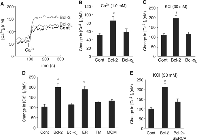
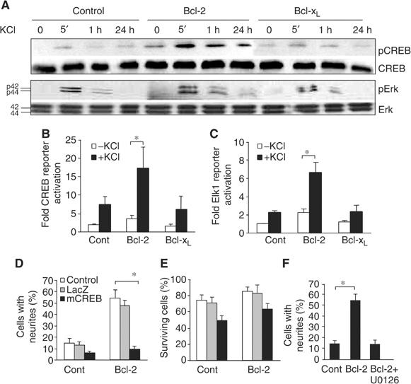
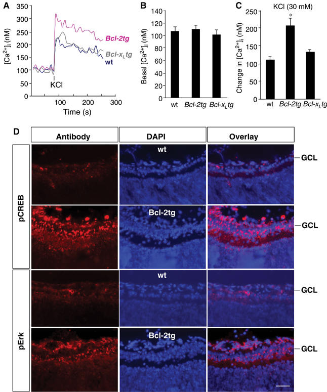
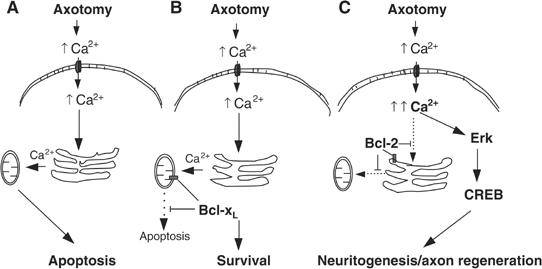
At a certain point in development, axons in the mammalian CNS undergo a profound loss of intrinsic growth capacity, which leads to poor regeneration after injury. Overexpression of Bcl-2 prevents this loss, but the molecular basis of this effect remains unclear. Here, we report that Bcl-2 supports axonal growth by enhancing intracellular Ca(2+) signaling and activating cAMP response element binding protein (CREB) and extracellular-regulated kinase (Erk), which stimulate the regenerative response and neuritogenesis. Expression of Bcl-2 decreases endoplasmic reticulum (ER) Ca(2+) uptake and storage, and thereby leads to a larger intracellular Ca(2+) response induced by Ca(2+) influx or axotomy in Bcl-2-expressing neurons than in control neurons. Bcl-x(L), an antiapoptotic member of the Bcl-2 family that does not affect ER Ca(2+) uptake, supports neuronal survival but cannot activate CREB and Erk or promote axon regeneration. These results suggest a novel role for ER Ca(2+) in the regulation of neuronal response to injury and define a dedicated signaling event through which Bcl-2 supports CNS regeneration.


Figures

Figure 1
Figure 1
Bcl-xL supports survival but not axon regeneration of postnatal RGCs. (A) Transverse retinal sections from wt, Bcl-xLtg, and Bcl-2tg mice were stained with TUNEL and GAP-43 antibody on day 1 after optic nerve injury. Asterisks indicate TUNEL-positive cells; arrows indicate the nerve fiber layer (NFL). TUNEL-positive cells are present and the NFL is absent in the retinal sections of wt mice but not Bcl-xLtg or Bcl-2tg mice. GCL, ganglion cell layer; IPL, inner plexiform layer. Scale bar, 100 μm. (B) Number of TUNEL-positive cells in retinal sections of wt, Bcl-xLtg and Bcl-2tg mice (n=4/group). (C) Longitudinal optic nerve sections from wt, Bcl-xLtg, and Bcl-2tg mice labeled with GAP-43 antibody 1–2 days after optic nerve injury. Arrows point to the crush site. Labeled axons in Bcl-xLtg mice remain anterior to the crush site, while those in Bcl-2tg mice grew robustly past the lesion site. Scale bar, 250 μm. (D) Retinal axon regrowth in retina–brain slice cocultures prepared from wt, Bcl-xLtg, and Bcl-2tg mice. Values are mean±s.d. *P<0.01 versus wt, two-tailed t-test.
Figure 2
Figure 2
Bcl-2 acts intrinsically in neurons to support neuritogenesis and axon regeneration. (A–C) RGCs were isolated from wt (A), Bcl-xLtg (B), and Bcl-2tg (C) mice, incubated for 5 days, and stained with calcein. Surviving RGCs from Bcl-2tg mice extended longer axons than those from wt or Bcl-xLtg mice. (D–F) Percentage of surviving RGCs (D), percentage of surviving RGCs with axons longer than 3 body lengths (E), and average length of the longest axon from each RGC (F) (n=5 cultures/group). RGCs in Bcl-xLtg and Bcl-2tg mice had similar survival rates, but Bcl-xLtg RGCs had shorter neurites. (G) Western blot analysis of Bcl-2 and Bcl-xL expression in stably transfected PC12 cells. (H, I) Percentage of surviving cells (H) and percentage of cells bearing neurites (I) (n=5 cultures/group). Bcl-xL- and Bcl-2-expressing PC12 cells had similar survival rates, but significantly fewer Bcl-xL-expressing cells had neurites. *P<0.01 versus wt, two-tailed t-test.
Figure 3
Figure 3
The growth-promoting effect of Bcl-2 is ER-dependent. (A) Schematic of DNA structures of Bcl-2, Bcl-xL, and Bcl-2 mutants. (B) Western blot analysis of PC12 cell lines stably transfected with Bcl-2ER, Bcl-2TM, and Bcl-2MOM. (C) Subcellular location of Bcl-2ER, Bcl-2MOM, and Bcl-xL shown by confocal microscopy. EGFP-Bcl-2ER (green) colocalizes with the ER marker calnexin (blue); EGFP-Bcl-2MOM and EGFP-Bcl-xL colocalize with the mitochondrial marker (Mito) cytochrome c (red). Scale bar, 4 μm. (D) Percentage of dying cells after treatment with staurosporine (left) and percentage that extended neurites (n=4 cultures/group). ER, Bcl-2ER; TM, Bcl-2TM; MOM, Bcl-2MOM. *P<0.05 versus wt, two-tailed t-test.
Figure 4
Figure 4
Bcl-2-, but not Bcl-xL-, expressing neurons display reduced ER Ca2+ content. (AC) Representative trace (A) and quantitative analysis of basal (B) and TG-induced [Ca2+]i (C), measured by Fura-2, in PC12 cells expressing a control (Cont), Bcl-2, or Bcl-xL plasmid. Measurement of TG-induced Ca2+ change was carried out in the absence of extracellular Ca2+. (D, E) TG-induced [Ca2+]i (D) and NGF-induced neurite outgrowth (E) in control (Cont), Bcl-2-, or Bcl-2+SERCA2b-expressing PC12 cells (n=4 cultures/group). (F) NGF-induced neurite outgrowth in control and Bcl-2-expressing PC12 cells treated with 0–10 mM BHQ (n=4/group). *P<0.01 versus control, two-tailed t-test.
Figure 5
Figure 5
Bcl-2 expression enhances intracellular Ca2+ signaling by reducing ER Ca2+ uptake. (AC) Representative trace (A) and quantitative analysis of extracellular Ca2+- (B) and KCl-induced changes in [Ca2+]i (C), measured by Fura-2 (n=4/group). PC12 cells stably transfected with control (Cont), Bcl-2, or Bcl-xL plasmids were incubated in Ca2+-free medium for over an hour; where indicated, Ca2+ (1.0 mM free extracellular Ca2+ final concentration) was added. Changes in [Ca2+]i were measured from its baseline to when the elevation of [Ca2+]i reached its peak. (D) Comparison of KCl-induced [Ca2+]i in cells expressing control (Cont), Bcl-2, Bcl-xL, Bcl-2ER (ER), Bcl-2TM (TM), and Bcl-2MOM (MOM) genes (n⩾3/group). (E) Measurement of KCl-induced changes in [Ca2+]i in cells expressing a control, Bcl-2, or Bcl-2+SERCA2b plasmid. *P<0.01 versus control, two-tailed t-test.
Figure 6
Figure 6
Bcl-2 activates CREB and Erk to promote neuritogenic response. (A) Western blot analysis of time-dependent phosphorylation of CREB and Erk in PC12 cells expressing control, Bcl-2, or Bcl-xL plasmids treated with KCl (30 mM). Antibodies recognizing the unphosphorylated forms of CREB and Erk served as controls. (B, C) CREB-dependent (B) and Erk-dependent (C) reporter gene activities in stably transfected PC12 cells in the presence or absence of KCl (30 mM) (n⩾4/group). (D–F) Neurite outgrowth (D, F) and neuronal survival (E) in control (Cont) and Bcl-2-expressing cells (Bcl-2) incubated with 1 ng/ml NGF (n=4/group). Cultures were incubated in the absence (control) or presence of either a viral vector carrying a LacZ reporter gene or dominant-negative CREB (mCREB) (D, E) or the MEK inhibitor U0126 (100 nM) (F). *P<0.01, two-tailed t-test.
Figure 7
Figure 7
Optic nerve injury in Bcl-2tg mice increases the intracellular Ca2+ response and activates CREB and Erk in RGCs. (A–C) Representative trace (A) and quantitative analysis of basal (B) and KCl-induced changes in [Ca2+]i (C), measured by Fura-2, in RGCs of wt, Bcl-2tg, and Bcl-xLtg mice (n⩾3/group). Changes in [Ca2+]i were measured from its baseline to when the elevation of [Ca2+]i reached its peak. Values are means±s.d. *P<0.01 versus wt, two-tailed t test. (D) Immunofluorescence staining for pCREB and pErk in retinal sections from adult wt and Bcl-2tg mice at day 1 after optic nerve crush. Scale bar, 50 μm.
Figure 8
Figure 8
Model of intracellular Ca2+ responses and subsequent signaling events in RGCs of wt, Bcl-xLtg, and Bcl-2tg mice. (A) Neural injury or axotomy results in an increase of extracellular Ca2+, a surge of Ca2+ influx, and elevation of [Ca2+]i. In neurons of wt mice, the excess intracellular Ca2+ is absorbed by the ER and translocated to the mitochondria, where it initiates apoptosis. (B) Bcl-xL expression, which is targeted to the mitochondria, prevents Ca2+-induced activation of the apoptotic signal and supports neuronal survival but does not affect ER Ca2+ uptake or the intracellular Ca2+ response to injury. (C) Expression of Bcl-2, however, is targeted primarily to the ER, where it reduces ER Ca2+ uptake and Ca2+ translocation to the mitochondria after axotomy, thereby preventing neuronal apoptosis. In addition, Bcl-2 reduces ER Ca2+ uptake, leading to greater elevation of [Ca2+]i after injury, thereby activating CREB and Erk and stimulating a neuritogenic response and axon regeneration.

Similar articles

Cited by

References

    1. Adams JP, Sweatt JD (2002) Molecular psychology: roles for the ERK MAP kinase cascade in memory. Annu Rev Pharmacol Toxicol 42: 135–163 - PubMed
    1. Berridge MJ (1998) Neuronal calcium signaling. Neuron 21: 13–26 - PubMed
    1. Chen DF, Jhaveri S, Schneider GE (1995) Intrinsic changes in developing retinal neurons result in regenerative failure of their axons. Proc Natl Acad Sci USA 92: 7287–7291 - PMC - PubMed
    1. Chen DF, Schneider GE, Martinou JC, Tonegawa S (1997) Bcl-2 promotes regeneration of severed axons in mammalian CNS. Nature 385: 434–439 - PubMed
    1. Cho K-S, Yang L, Lu B, Ma HF, Huang X, Pekny M, Chen DF (2005) Re-establishing the regenerative potential of central nervous system axons in postnatal mice. J Cell Sci (in press) - PMC - PubMed

Publication types

MeSH terms