. 2005 Mar 9;24(5):1046-56.
doi: 10.1038/sj.emboj.7600580. Epub 2005 Feb 17.

Maintenance of structure and function of mitochondrial Hsp70 chaperones requires the chaperone Hep1

Affiliations

Maintenance of structure and function of mitochondrial Hsp70 chaperones requires the chaperone Hep1

Martin Sichting et al. EMBO J. .

Abstract

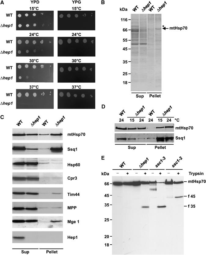
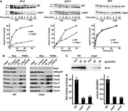
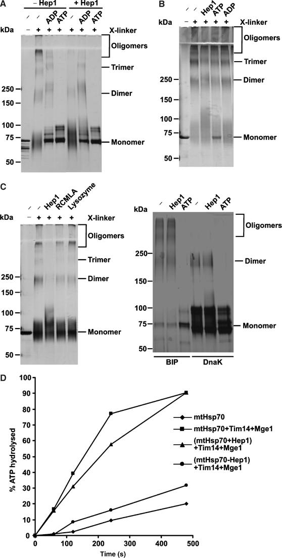
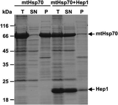
Hsp70 chaperones mediate folding of proteins and prevent their misfolding and aggregation. We report here on a new kind of Hsp70 interacting protein in mitochondria, Hep1. Hep1 is a highly conserved protein present in virtually all eukaryotes. Deletion of HEP1 results in a severe growth defect. Cells lacking Hep1 are deficient in processes that need the function of mitochondrial Hsp70s, such as preprotein import and biogenesis of proteins containing FeS clusters. In the mitochondria of these cells, Hsp70s, Ssc1 and Ssq1 accumulate as insoluble aggregates. We show that it is the nucleotide-free form of mtHsp70 that has a high tendency to self-aggregate. This process is efficiently counteracted by Hep1. We conclude that Hep1 acts as a chaperone that is necessary and sufficient to prevent self-aggregation and to thereby maintain the function of the mitochondrial Hsp70 chaperones.


Figures

Figure 1
Figure 1
Hep1 interacts with mtHsp70. (A, B) Co-isolation of Hep1 with His-tagged mtHsp70. Isolated mitochondria from WT or from a yeast strain expressing Hsp70 with a hexahistidinyl tag were solubilized in Triton X-100. (A) Mitochondrial lysates were passed over a NiNTA column. Bound material was eluted and analysed by SDS–PAGE and Coomassie staining. Tim44, Mge1 and Hep1 were identified by mass spectrometry. (B) Mitochondrial lysates were incubated with NiNTA beads and the beads were isolated. Total proteins, T (10%), unbound material, FT (10%) and bound material, B (100%), were analysed by SDS–PAGE and immunodecoration for the indicated proteins. (C) Co-immunoprecipitation of Hep1 with mtHsp70. WT mitochondria were either depleted of ATP by incubation with apyrase and oligomycin (−ATP) or incubated with an ATP regenerating system (+ATP) for 10 min. Following lysis in digitonin, mitochondrial proteins were incubated with antibodies against mtHsp70 or with preimmune serum (PI). Immunoprecipitates were analysed by SDS–PAGE and immunodecoration with antibodies against the indicated proteins. The total and supernatant (SN) represent 20% of the material of the precipitate (P). (D) Hep1 can be crosslinked to mtHsp70. Mitochondria from WT cells and cells expressing His-tagged mtHsp70 were treated as in (C) and then subjected to crosslinking with DSG. Control samples without crosslinking reagent and aliquots after crosslinking were directly subjected to SDS–PAGE (T); the other aliquots were solubilized and incubated with NiNTA beads. Bound material was eluted and subjected to SDS–PAGE. Proteins were analysed by Western blotting using antibodies directed against Hep1.
Figure 2
Figure 2
Hep1 does not increase the ATPase activity and the nucleotide exchange rate of mtHsp70. (A) Coomassie-stained gel of purified proteins Hep1-His6 and mtHsp70. (B) The ATPase activity of mtHsp70 was determined by the rate of conversion of 32P-ATP to ADP and 32P. Samples containing the indicated purified proteins were incubated for the indicated times, subjected to thin layer chromatography and the percentage of hydrolysed ATP was quantified by phosphoimaging. A fusion protein of MBP and Tim14 was used (Tim14). (C) Preformed complex of mtHsp70 with 32P-ATP was incubated at 30°C with excess of unlabelled ATP in the presence of Hep1, Mge1 or both Mge1 and Hep1 as indicated. The fraction of hydrolysed 32P-ATP was determined at the indicated time points as in (B).
Figure 3
Figure 3
Deletion of HEP1 leads to aggregation of mitochondrial Hsp70 proteins. (A) Drop dilution test of WT and a HEP1 deletion strain (Δhep1). Dilutions, 10-fold, were plated on YPD and YPG plates and incubated at the indicated temperatures. (B, C) Aggregation of mtHsp70 in cells lacking Hep1. Mitochondria isolated from WT and Δhep1 cells were lysed with 1% digitonin and aggregated material was pelleted by centrifugation (Pellet). The SN fraction (Sup) was precipitated with TCA. Pellets and Sups were analysed by SDS–PAGE and Coomassie staining (B) or immunodecoration with antibodies against mtHsp70, Ssq1 and the other indicated proteins (C). (D) Aggregation of Hsp70 proteins depends on the growth temperature of cells. WT and Δhep1 cells were grown at 15°C and 24°C. Then, mitochondria were isolated and the aggregation state of mtHsp70 and Ssq1 was tested as above. Equal amounts of protein from the Pellet and Sup fraction were analysed. (E) Increased protease sensitivity of mtHsp70 in the absence of Hep1. Mitochondria were isolated from WT, from Δhep1 cells and from temperature-sensitive mutants, ssc1–3 and ssc1–2, grown at permissive temperature. Mitochondria were lysed in the presence of Triton X-100 and treated with trypsin for 5 min on ice. Samples were analysed by SDS–PAGE and immunodecoration with antibodies against full-length mtHsp70. The obtained fragments are indicated: f 45, N-terminal fragment; f 35, C-terminal fragment.
Figure 4
Figure 4
Cells lacking Hep1 are defective in import of mitochondrial preproteins and in the biogenesis of FeS proteins. (A) Mitochondria isolated from Δhep1 cells and from WT were incubated with radiolabelled preproteins. The substrates pF1β (subunit β of the F1Fo-ATPase) and pJac1 of the TIM23 complex and the substrate of the TIM22 complex, ATP/ADP carrier (AAC), were used as preproteins. Mitochondria were treated with proteinase K, reisolated and analysed by SDS–PAGE and autoradiography. The mature forms of the proteins were quantified. Import into WT mitochondria at the longest time point was set to 100%. p, precursor form; m, mature form. (B) Hep1 is not part of the TIM23 complex. Mitochondria were isolated from WT cells (left panel) and cells lacking Hep1 (right panel). After solubilization with digitonin, the lysate was subjected to immunoprecipitation with antibodies against Tim17, Tim16 and with preimmune serum (PI). Samples were analysed by SDS–PAGE and immunodecoration with antibodies against the indicated proteins of the TIM23 complex and Hep1. ‘Total' and ‘SNs' represent 20% of material present in ‘Pellets'. (C) Mitochondrial proteins were analysed by SDS–PAGE and immunodecoration with antibodies against aconitase. (D) The activities of SDH and malate dehydrogenase (MDH) were measured in mitochondria from WT, Δhep1 and Δssq1 cells. The SDH activities are given per mg of mitochondrial protein (left panel) and relative to the activity of malate dehydrogenase (right panel). The ratio obtained for WT mitochondria was set as 1. Error bars show the standard deviation of three independent experiments.
Figure 5
Figure 5
Hep1 prevents self-aggregation of mtHsp70. (A) Hep1 inhibits aggregation of mtHsp70. Purified mtHsp70 was incubated for 10 min at 30°C in the absence or presence of the indicated nucleotides and Hep1. The samples were subjected to crosslinking with glutaraldehyde and analysed by SDS–PAGE (6–14% gradient gel) and silver staining. (B) Self-aggregation of mtHsp70 is not reversible. MtHsp70 was first incubated for 10 min at 30°C to allow oligomerization and then kept for 10 min in the presence of nucleotides and Hep1 as indicated. Further treatment as in (A). (C) Prevention of self-aggregation is specific for Hep1 and mtHsp70. Left panel: mtHsp70 (left panel) in the presence of lysozyme or RCMLA and purified BiP and DnaK (right panel) were treated and analysed as in (A). (D) Hep1 keeps mtHsp70 functional. The ATPase activity of mtHsp70 in the presence of the indicated proteins was determined as in Figure 2B. Where indicated, mtHsp70 was first preincubated for 30 min at 30°C either in the absence (Hsp70−Hep1) or presence (Hsp70+Hep1) of equimolar amounts of Hep1.
Figure 6
Figure 6
Hep1 prevents aggregation of mtHsp70 in E. coli cells. MtHsp70 was expressed in E. coli either alone (mtHsp70) or together with a His-tagged form of Hep1 (mtHsp70+Hep1). Both proteins lacked their matrix targeting sequences. Following induction of protein expression cells were lysed and one aliquot was removed (total, T). The other aliquot was separated by centrifugation into insoluble protein (P) and soluble protein fractions (SN). Samples were analysed by SDS–PAGE and Coomassie staining.

Similar articles

Cited by

References

    1. Angelidis CE, Lazaridis I, Pagoulatos GN (1999) Aggregation of hsp70 and hsc70 in vivo is distinct and temperature-dependent and their chaperone function is directly related to non-aggregated forms. Eur J Biochem 259: 505–512 - PubMed
    1. Azem A, Oppliger W, Lustig A, Jeno P, Feifel B, Schatz G, Horst M (1997) The mitochondrial hsp70 chaperone system. Effect of adenine nucleotides, peptide substrate, and mGrpE on the oligomeric state of mhsp70. J Biol Chem 272: 20901–20906 - PubMed
    1. Bateman A, Coin L, Durbin R, Finn RD, Hollich V, Griffiths-Jones S, Khanna A, Marshall M, Moxon S, Sonnhammer EL, Studholme DJ, Yeats C, Eddy SR (2004) The Pfam protein families database. Nucleic Acids Res 32, (database issue): D138–D141 - PMC - PubMed
    1. Baumann F, Milisav I, Neupert W, Herrmann JM (2000) Ecm10, a novel hsp70 homolog in the mitochondrial matrix of the yeast Saccharomyces cerevisiae. FEBS Lett 487: 307–312 - PubMed
    1. Benaroudj N, Triniolles F, Ladjimi MM (1996) Effect of nucleotides, peptides, and unfolded proteins on the self-association of the molecular chaperone HSC70. J Biol Chem 271: 18471–18476 - PubMed

Publication types

MeSH terms