. 2005 Mar 9;24(5):952-62.
doi: 10.1038/sj.emboj.7600579. Epub 2005 Feb 17.

A receptor domain controls the intracellular sorting of the ferrichrome transporter, ARN1

Affiliations

A receptor domain controls the intracellular sorting of the ferrichrome transporter, ARN1

Youngwoo Kim et al. EMBO J. .

Abstract

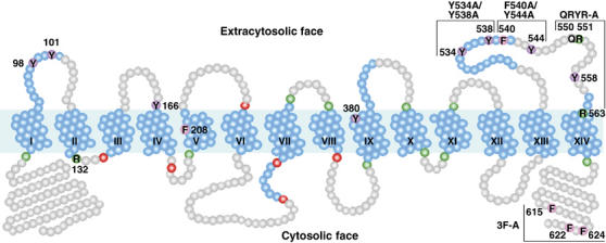
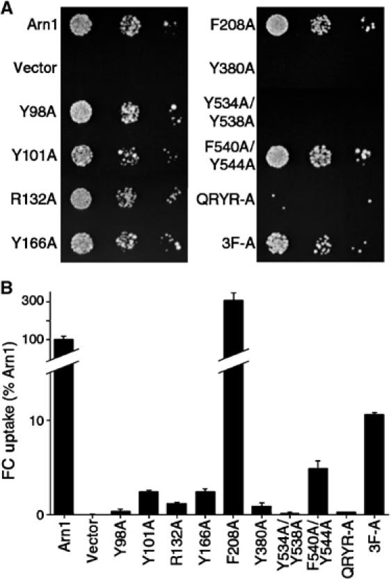
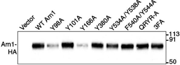
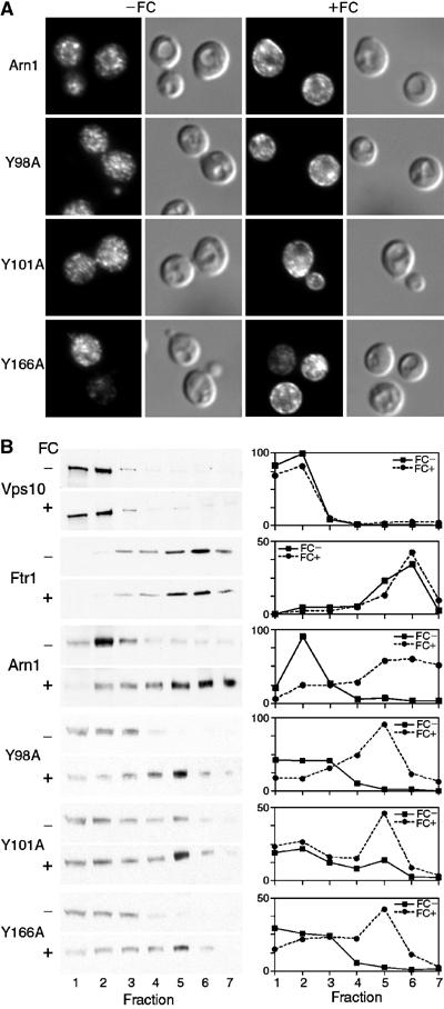
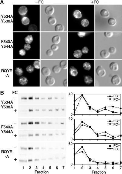
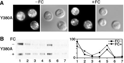
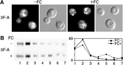
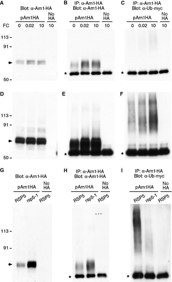
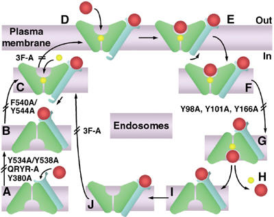
The Saccharomyces cerevisiae transporter Arn1p takes up the ferric-siderophore ferrichrome, and extracellular ferrichrome dramatically influences the intracellular trafficking of Arn1p. In the absence of ferrichrome, Arn1p sorts directly to the endosomal compartment. At low concentrations of ferrichrome, Arn1p stably relocalizes to the plasma membrane, yet little to no uptake of ferrichrome occurs at these low concentrations. At higher concentrations of ferrichrome, Arn1p cycles between the plasma membrane and endosome. Arn1p contains two binding sites for ferrichrome: one site has an affinity similar to the K(T) for transport, but the second site has a much higher affinity. Here we report that this high-affinity binding site lies within a unique extracytosolic, carboxyl-terminal domain. Mutations within this domain lead to loss of ferrichrome binding and uptake activities and missorting of Arn1p, including a failure to relocalize to the plasma membrane in the presence of ferrichrome. Mutation of phenylalanine residues in the cytosolic tail of Arn1p also lead to missorting, but without defects in ferrichrome binding. We propose that the carboxyl terminus of Arn1p contains a receptor domain that controls the intracellular trafficking of the transporter.


Figures

Figure 1
Figure 1
Predicted topology and mutagenesis of Arn1p. TMDs predicted from TMHMM and TMpred are numbered and shown coiled in blue, with additional potential helical regions predicted from HMMTOP and the structure of GlpT also in blue. Basic residues anchoring TMDs are shown in green, and acidic residues anchoring TMDs are shown in red. The region spanning the first 12 TMDs exhibits homology to other MFS family members. Mutated residues are numbered.
Figure 2
Figure 2
Impaired growth on FC and defects in uptake of FC due to mutations in Arn1p. A strain (CWY121) bearing deletions of all four Arn transporters and defective in ferrous iron uptake (fet3Δ) was transformed with low-copy-number plasmids containing the wild-type ARN1 allele, the indicated mutant arn1 alleles, or the empty vector. (A) Impaired growth on FC. Transformed strains were plated in serial 10-fold dilutions on medium containing 10 μM FC and incubated for 3 days at 30°C prior to imaging. (B) Defective uptake of FC. Transformed strains were grown in iron-poor medium to a density of 1 × 10⁷ cells/ml, cells were harvested, and the uptake of [55Fe]FC was measured. FC uptake activity was normalized to the strain transformed with the wild-type allele. Assays were performed in duplicate, experiments were replicated three times, and a representative experiment is shown.
Figure 3
Figure 3
Expression levels of wild-type and mutant alleles of Arn1p. The strain YPH499 was transformed with the wild-type and the mutant alleles of ARN1 in the low-copy number plasmid pRS316 and grown in iron-poor medium to induce expression of Arn1p. Cells were subjected to glass bead lysis and membranes were isolated. Identical quantities of membrane protein were analyzed by SDS–PAGE and Western blotting using antibodies against the HA epitope to detect alleles of Arn1p-HA. Molecular weight standards are indicated in kDa.
Figure 4
Figure 4
Relocalization to the plasma membrane of wild-type and mutant Arn1p after exposure to FC. (A) Plasma membrane relocalization by indirect immunofluorescence. The strain YPH499 was transformed with the wild-type and the indicated mutant alleles of Arn1p in the low-copy number plasmid pRS316 and grown in iron-poor medium to induce expression of Arn1p. Cells were divided and grown for an additional hour in the absence (−FC) or presence (+FC) of 100 nM FC prior to fixation and preparation for indirect immunofluorescence microscopy. Mouse monoclonal HA.11 was the primary antibody and Cy-3-conjugated donkey anti-mouse was the secondary antibody. Images are in pairs with fluorescence on the left and DIC on the right. (B) Redistribution of wild-type and mutant Arn1p to fractions containing plasma membranes after exposure to FC. The strain YPH499 FET3-HA FTR1-myc was transformed with the wild-type and the indicated mutant alleles of Arn1p in the low-copy number plasmid pRS316 and grown in the absence (FC−) or presence (FC+) of 100 nM FC as described above. Cells were harvested, membranes were collected and separated on sucrose step gradients, and the fractions were subjected to SDS–PAGE and Western blotting. Late Golgi vesicles were detected using an antibody directed against Vps10p, plasma membranes were detected using an anti-myc antibody directed against Ftr1p-myc, and HA-tagged Arn1p alleles were detected using anti-HA antibody. HA-tagged Fet3p (not shown) served as an internal loading control. Western blots are on the left, in pairs, and densitometric quantitation is shown on the right. Densitometry is reported in arbitrary units, which are proportional to the intensity of the bands on Western blots.
Figure 5
Figure 5
Loss of FC-induced plasma membrane relocalization in Arn1p mutants with substitutions in the last extracytosolic loop domain. Indirect immunofluorescence (A) and membrane fractionation (B) were performed as in Figure 4 using cells transformed with the indicated Arn1p alleles containing mutations in the last predicted extracytosolic loop between TMDs XIII and XIV.
Figure 6
Figure 6
Localization of mutant allele Y380A to the plasma membrane in the absence of FC. Indirect immunofluorescence (A) and membrane fractionation (B) were performed as in Figure 4 using cells transformed with the Arn1p mutant Y380A, which contained a mutation in the predicted extracytosolic terminus of TMD IX.
Figure 7
Figure 7
Impaired FC-induced relocalization to the plasma membrane in an Arn1p mutant with substitutions in the carboxyl-terminal cytosolic tail. Indirect immunofluorescence (A) and membrane fractionation (B) were performed as in Figure 4 using cells transformed with the Arn1p mutant 3F-A containing substitutions of three phenylalanine residues in the predicted carboxyl-terminal cytosolic tail.
Figure 8
Figure 8
FC-induced, Rsp5-dependent ubiquitination of Arn1p. Strains YPH499 (A–F), LHY23 (G–I, rsp5-1), and LHY291 (G–I, RSP5) were transformed with either pMET Arn1-HA (pArn1HA) or the empty vector pRS416 (no HA) and Yep105, which expresses myc-tagged ubiquitin from the CUP1 promoter. Transformed strains were grown in synthetic complete medium without methionine and with 100 μM copper to induce expression of Arn1-HA and myc-ubiquitin, respectively. FC (μM) was added at the indicated concentrations for 2 h (A–F) or cultures were shifted to 37°C with the addition of 5 μM FC for 2 h (G–I). Then, an equivalent number of cells was harvested from each culture. Cells were lysed and an aliquot was subjected to Western blotting with anti-HA antibody (A, D, G). Membranes were purified from the remaining lysate, detergent solubilized, and subjected to immunoprecipitation with anti-HA antibody. Immunoprecipitates were then subjected to Western blotting using anti-HA antibodies to detect Arn1-HA (B, E, H) or anti-myc antibodies to detect myc-ubiquitin (C, F, I). Panels D–F are an overexposure of blots depicted in panels A–C to more clearly demonstrate ubiquitinated species. Panel I is approximately five-fold overexposed with respect to panel H. The arrow indicates unmodified Arn1-HA. The asterisk indicates immunoglobulin heavy chain.
Figure 9
Figure 9
Proposed mechanism for trafficking of Arn1p and uptake of FC. Apo-Arn1p is sorted to recycling endosomes (A). FC enters the endosome through fluid-phase endocytosis and, upon binding to the receptor site (B), triggers a conformational change in the cytosolic tail and stabilization of the outward-open conformation (C), then proton binding and relocalization to the plasma membrane (D). Upon binding of a second molecule of FC in the transport site (E), Arn1p undergoes endocytosis (F), which triggers a shift to the inward-open conformation (G), wherein FC and the proton are released (H). FC bound to the receptor site (I) can then again trigger stabilization of the outward-open conformation (J) and conformational change in the cytosolic tail (C). The transport domains (green) and receptor domain (blue) of Arn1p, iron-bound FC (red), and proton (yellow) are shown. Double bars mark the steps that were inhibited by the indicated mutations.

Similar articles

Cited by

References

    1. Abramson J, Smirnova I, Kasho V, Verner G, Iwata S, Kaback HR (2003a) The lactose permease of Escherichia coli: overall structure, the sugar-binding site and the alternating access model for transport. FEBS Lett 555: 96–101 - PubMed
    1. Abramson J, Smirnova I, Kasho V, Verner G, Kaback HR, Iwata S (2003b) Structure and mechanism of the lactose permease of Escherichia coli. Science 301: 610–615 - PubMed
    1. Aridor M, Traub LM (2002) Cargo selection in vesicular transport: the making and breaking of a coat. Traffic 3: 537–546 - PubMed
    1. Askwith C, Eide D, Van Ho A, Bernard PS, Li L, Davis-Kaplan S, Sipe DM, Kaplan J (1994) The FET3 gene of S. cerevisiae encodes a multicopper oxidase required for ferrous iron uptake. Cell 76: 403–410 - PubMed
    1. Bagnat M, Chang A, Simons K (2001) Plasma membrane proton ATPase pma1p requires raft association for surface delivery in yeast. Mol Biol Cell 12: 4129–4138 - PMC - PubMed

MeSH terms