. 2005 Mar;114(3):418-27.
doi: 10.1111/j.1365-2567.2005.02090.x.

Lupus autoantibodies to native DNA preferentially bind DNA presented on PolIV

Affiliations

Lupus autoantibodies to native DNA preferentially bind DNA presented on PolIV

Sanjeev Kumar et al. Immunology. 2005 Mar.

Abstract

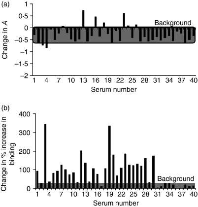
While immunoglobulin G (IgG) antibodies to double-stranded (ds)DNA are serological markers of systemic lupus erythematosus (SLE), not all antibodies to DNA (anti-DNA) are able to cause tissue damage to a similar extent. It has been proposed that anti-DNA-induced renal damage could be linked to differences in the fine specificity of the antibodies. In an attempt to gain insight into their fine binding properties, we investigated the cross-reactivity of two human lupus monoclonal IgG anti-dsDNA (B3 and RH14) to a recently described Escherichia coli PolIV (a DNA polymerase). These autoantibodies possess distinct pathogenic properties in severe combined immunodeficient (SCID) mice. Although both antibodies cause proteinuria, only RH14 induces early histological features of lupus nephritis. Both RH14 and B3 bound PolIV; however, they exhibited a marked difference in their reactivity to the PolIV-dsDNA complex. Alhough RH14 exhibited significant activity to the complex, the binding of B3 to PolIV complexed with dsDNA was almost abolished. Furthermore, there was a significant difference in the way the lupus sera recognized naked dsDNA and that presented on PolIV. Although 67% of lupus sera bound naked dsDNA, approximately 90% of these sera (93% calf thymus DNA; 90% synthetic oligonucleotide) reacted to the complex when dsDNA was presented on PolIV. Thus, the IgG anti-dsDNA likely to exist in lupus patients may be distinguished into those that recognize dsDNA in the context of PolIV and those which do not. This difference in binding ability may help to distinguish those dsDNA antibodies that are more pathogenic.


Figures

Figure 1
Figure 1
(a) Sodium dodecyl sulphate–polyacrylamide gel electrophoresis (SDS–PAGE) gel profile of the purified PolIV. The gel was stained with Coomassie Blue. PolIV resolves at 40 500 molecular weight (MW) – the correct size of the molecule. Lane 1, MW markers; and lane 2, purified PolIV (7·5 µg). (b)Electrophoretic mobility shift assay in a non-denaturing PAGE system, demonstrating binding of purified PolIV to a primer-template terminus. Relative positions of the unbound oligonucleotide and of the protein controls are indicated, compared to the DNA–protein complex. Migration of the small, negatively charged oligonucleotide is severely retarded in the presence of PolIV, owing to the positive charge on the protein (theoretical pI of 9·28). In the absence of oligonucleotide, the positive charge on PolIV prevents migration of the protein into the gel. The presence of both DNA and protein in the complex is confirmed by duplicate staining in Coomassie Blue (i) and ethidium bromide (ii). Lane 1, oligonucleotide alone (control, amount equivalent to oligonucelotide present in lane 2); lane 2, PolIV–oligonucleotide complex (6 µg of protein); and lane 3, PolIV alone (control, 6 µg of protein).
Figure 2
Figure 2
(a) Binding of the 35mer biotinylated oligonucleotides in their single-stranded (ss) or double-stranded (ds) forms, to PolIV alone or to PolIV-preblocked calf thymus dsDNA, in a solid phase. (b) Binding of the monoclonal human dsDNA antibodies RH14 and B3 to PolIV or PolIV + ds/ss DNA or (c) to poly-l-lysine or poly-l-lysine + dsDNA, in a solid phase. (d) Binding of calf thymus ds/ssDNA-preblocked RH14 and B3 to PolIV or PolIV + ds/ss DNA, in a solid phase.
Figure 3
Figure 3
Binding of 30 sera from the patients with lupus (numbers 1–15 with nephritis; 16–30, without nephritis) and of 10 sera from normal healthy individuals (numbers 31–40) to naked calf thymus double-stranded (ds)DNA in a traditional enzyme-linked immunosorbent assay (ELISA). The highest absorbance (A) at 405 nm (0·763) recorded for the normal sera, was used as the background and the lower limit to determine the anti-naked dsDNA activity of lupus sera.
Figure 4
Figure 4
Binding of 30 sera from the patients with lupus (numbers 1–15 with or numbers 16–30 without lupus nephritis) and of 10 sera from normal healthy individuals (numbers 31–40) to a PolIV–calf thymus double-stranded (ds)DNA complex (a) or to a PolIV–oligonucleotide dsDNA complex (c), and the percentage increase in their binding to these complexes, in relation to their binding to PolIV alone, following the presentation of calf thymus (b)or oligonucleotide (d)dsDNA on PolIV.
Figure 5
Figure 5
A comparison of the binding of 30 sera from patients with lupus (numbers 1–15 with or numbers 16–30 without lupus nephritis) and of 10 sera from normal healthy individuals (numbers 31–40), to naked double stranded (ds)DNA and to a PolIV–dsDNA complex. (a) The difference in binding of the serum autoantibodies to naked or to PolIV-complexed calf thymus dsDNA was determined by using the following equation: Change in A = (APolIV+dsDNAAPolIV) − (AdsDNAAplate background). (b)The difference in percentage increase in the binding of the serum autoantibodies to calf thymus or to synthetic oligonucleotide dsDNA complexed with PolIV, was determined by using the following equation: Change in percentage increase in binding = (% increase in binding to PolIV–calf thymus dsDNA) − (% increase in binding to PolIV-oligonucleotide dsDNA).

Similar articles

Cited by

References

    1. Swaak AJG, Aarden LA, Statius van Eps LW, Feltkamp TE. Anti-dsDNA and complement profiles as prognostic guides in systemic lupus erythematosus. Arthritis Rheum. 1979;22:226–35. - PubMed
    1. Isenberg DA, Garton M, Reichlin MW, Reichlin M. Long-term follow-up of autoantibody profiles in black female lupus patients and clinical comparison with Caucasian and Asian patients. Br J Rheumatol. 1997;36:229–33. - PubMed
    1. Okamura M, Kanayama Y, Amastu K, Negoro N, Kohda S, Takeda T, Inoue T. Significance of enzyme linked immunosorbant assay (ELISA) for antibodies to double stranded and single stranded DNA in patients with lupus nephritis: correlation with severity of renal histology. Ann Rheum Dis. 1993;52:14–20. - PMC - PubMed
    1. Isenberg DA, Shoenfeld Y, Walport M, et al. Detection of cross-reactive anti-DNA antibody idiotypes in the serum of systemic lupus erythematosus patients and of their relatives. Arthritis Rheum. 1985;28:999–1007. - PubMed
    1. Cabral AR, Alarcon-Segovia D. Autoantibodies in systemic lupus erythematosus. Curr Opin Rheumatol. 1997;9:387–92. - PubMed

Publication types