

Case-controlled clinical and histopathological study of conjunctivochalasis
- PMID: 15722309
- PMCID: PMC1772548
- DOI: 10.1136/bjo.2004.051144
Case-controlled clinical and histopathological study of conjunctivochalasis
Abstract
Background/aims: Conjunctivochalasis, a secondary cause of the watery eye, is frequently seen in the older age group as an elevation of the bulbar conjunctiva lying along the lateral or central lower lid margin. A prospective, interventional, case-controlled clinical and histopathological study was conducted. The relevant features of 18 patients (29 eyes) who had their conjunctivochalasis resected as part of the surgical management of their watery eye syndrome were examined. In the control group, tissue was obtained from an age matched series of 24 normal subjects undergoing routine cataract surgery.
Methods: 24 controls (24 specimens) and 18 patients (29 specimens) had conjunctival strip biopsies, taken from the usual lid margin level bulbar conjunctiva in line with the inferior limbus (controls), and the clinically apparent conjunctivochalasis (patients). These were submitted for histological study.
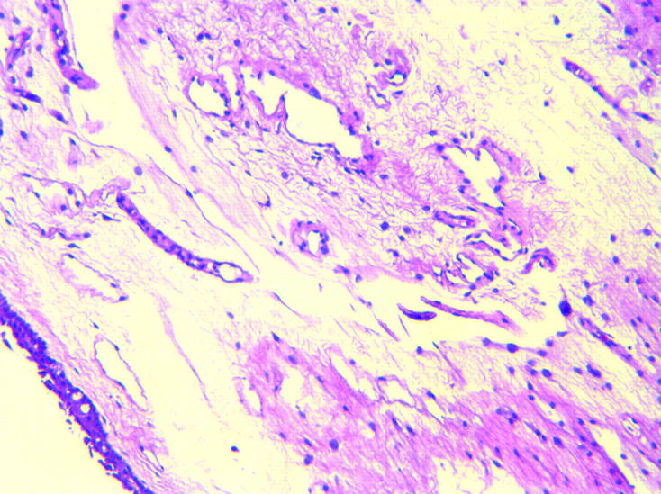
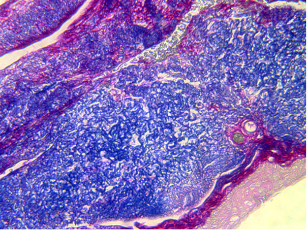
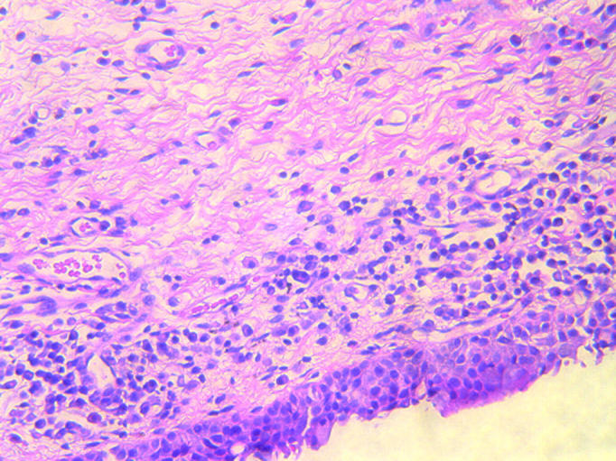
Results: 23 of 24 control sections demonstrated normal conjunctival variation. Four of 29 patient specimens demonstrated a chronic non-granulomatous conjunctivitis, while three eyes of the patient group (two patients) demonstrated features of elastosis. Of the four patients who had the inflammatory infiltrates, three had functional nasolacrimal duct obstructions (FNLDOs) and one had a primary acquired nasolacrimal duct obstruction (PANDO). Of the two patients who had elastosis, one had an FNLDO and the other had normal lacrimal drainage and was Jones 1 positive.
Conclusion: Six of 18 patients--that is, seven of 29 specimens of conjunctivochalasis demonstrated signs of elastosis or of chronic non-granulomatous inflammation. Clinically, patients had a spectrum of aetiologies of their watery eye syndrome.
Figures
References
-
- Braunschweig P. Uber Faltenbildung der Conjunctiva bulbi. Klin Monatsbl Augenheilkd 1921;66:123–4.
-
- Hughes WL. Conjunctivochalasis. Am J Ophthalmol 1942;25:48–51.
-
- Duke-Elder S. Conjunctival hyperplasia. System of ophthalmology. Vol XIII: The ocular adnexa. London: Kimpton, 1974:609.
-
- Liu D. Conjunctivochalasis. A cause of tearing and its management. Ophthalmic Plast Reconstr Surg 1986;2:25–8. - PubMed
-
- Meller D, Tseng SCG. Conjunctivochalasis: literature review and possible pathophysiology. Surv Ophthalmol 1998;43:225–32. - PubMed
MeSH terms
LinkOut - more resources
Full Text Sources