

Optical coherence tomography in photodynamic therapy for subfoveal choroidal neovascularisation secondary to age related macular degeneration: a cross sectional study
- PMID: 15722312
- PMCID: PMC1772543
- DOI: 10.1136/bjo.2004.043364
Optical coherence tomography in photodynamic therapy for subfoveal choroidal neovascularisation secondary to age related macular degeneration: a cross sectional study
Abstract
Aims: To introduce new terminology and validate its reliability for the analysis of optical coherence tomography (OCT) scans, compare clinical detection of cystoid macular oedema (CMO) and subretinal fluid (SRF) with OCT findings, and to study the effect of photodynamic therapy (PDT) on the foveal morphology.
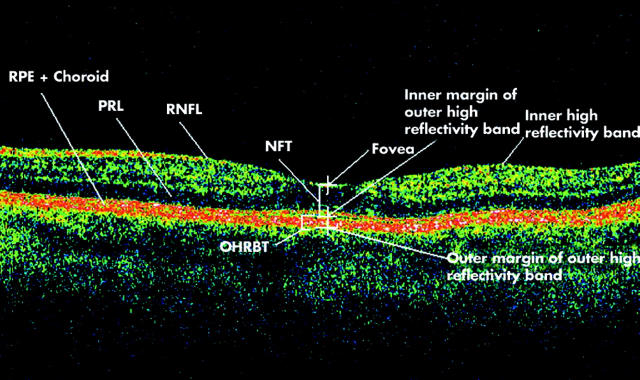
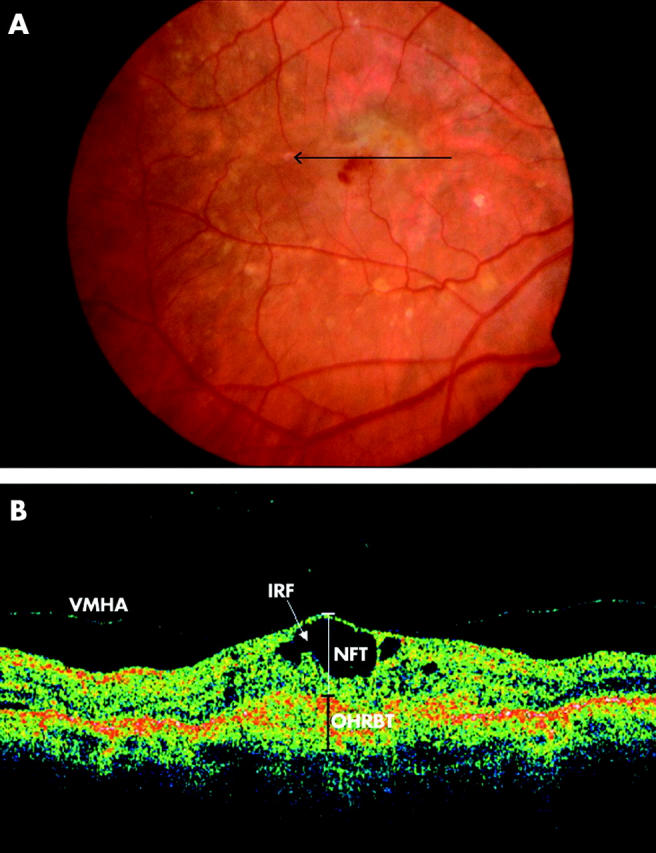
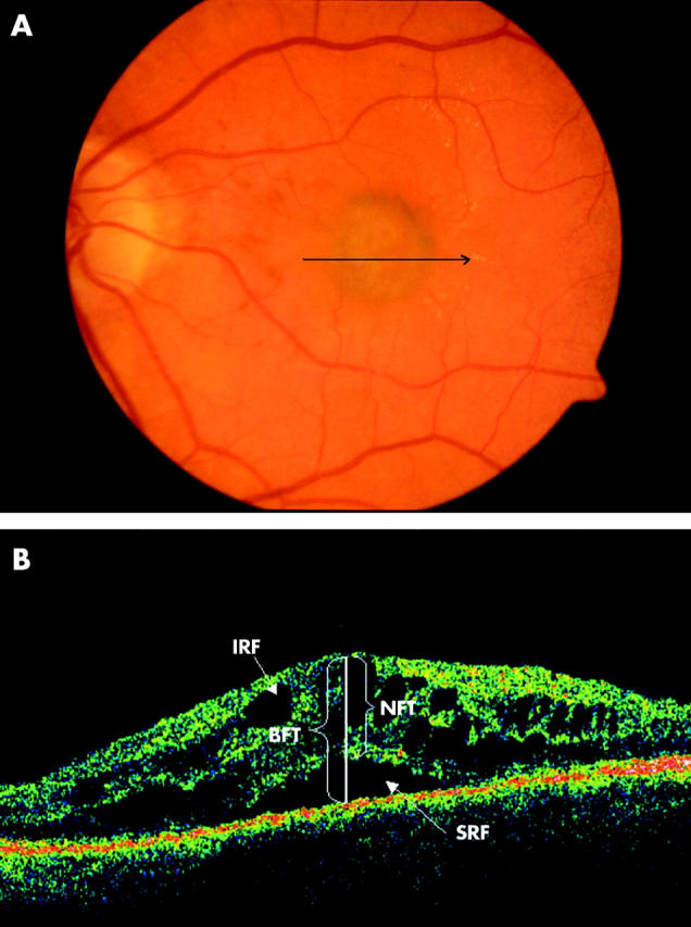
Methods: Patients with subfoveal, predominantly classic choroidal neovascularisation (CNV) secondary to age related macular degeneration (AMD) undergoing PDT were evaluated with refraction protocol best corrected logMAR visual acuity (VA), slit lamp biomicroscopy, stereoscopic fluorescein angiography (FFA), and OCT. New terminologies introduced to interpret the OCT scans were: neuroretinal foveal thickness (NFT), bilaminar foveal thickness (BFT), outer high reflectivity band thickness (OHRBT), intraretinal fluid (IRF), subretinal fluid (oSRF), and vitreomacular hyaloid attachment (VMHA).
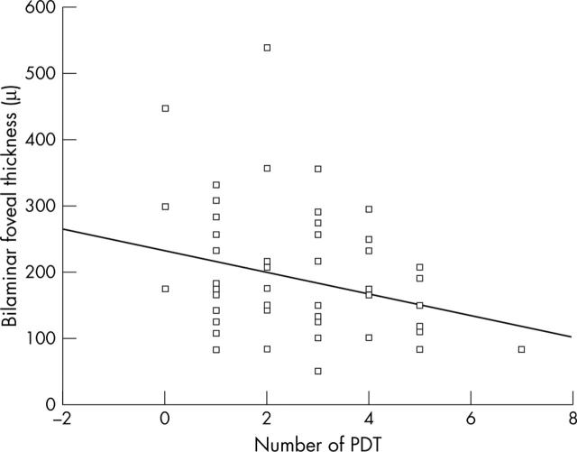
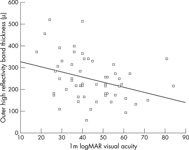
Results: Fifty six eyes of 53 patients were studied. VA was better in eyes with a thinner outer high reflectivity band (OHRBT) (p = 0.02) and BFT (p = 0.05). BFT was less in eyes that had undergone a greater number of PDT treatments (p = 0.04). There was poor agreement between OCT and clinical examination in the detection of CMO and subretinal fluid (kappa = 0.289 and kappa = 0.165 respectively). To validate the interpretation and measurements on OCT, two groups of 20 scans were analysed by two independent observers. There was good agreement between the observers in the detection of IRF, oSRF, and VMHA (p<0.001). Measurements of NFT and BFT had a high reproducibility, and of OHRBT reproducibility was low.
Conclusions: New terminology has been introduced and tested. OCT appears to be superior to clinical examination and FFA in the detection of CMO. In this study, better vision was associated with a thinner OHRBT and/or the absence of SRF giving insight into the biological effect of PDT.
Figures
Comment in
-
Optical coherence tomography in photodynamic therapy.Br J Ophthalmol. 2005 Jul;89(7):928-9. doi: 10.1136/bjo.2005.071092. Br J Ophthalmol. 2005. PMID: 15965188 Free PMC article. No abstract available.
-
Optical coherence tomography of the vitreomacular interface in photodynamic therapy.Br J Ophthalmol. 2005 Jul;89(7):929; author reply 929. Br J Ophthalmol. 2005. PMID: 15965190 Free PMC article. No abstract available.
Similar articles
-
Optical coherence tomography analysis of a randomized study combining photodynamic therapy with intravitreal triamcinolone.Graefes Arch Clin Exp Ophthalmol. 2008 Feb;246(2):245-54. doi: 10.1007/s00417-007-0642-1. Epub 2007 Aug 3. Graefes Arch Clin Exp Ophthalmol. 2008. PMID: 17674020 Clinical Trial.
-
Optical coherence tomography analysis of bilateral end-stage choroidal neovascularization where one eye is treated with photodynamic therapy.Clin Exp Ophthalmol. 2007 Jan-Feb;35(1):13-7. doi: 10.1111/j.1442-9071.2006.01385.x. Clin Exp Ophthalmol. 2007. PMID: 17300565
-
Intravitreal bevacizumab for previously treated choroidal neovascularization from age-related macular degeneration.Retina. 2007 Apr-May;27(4):432-8. doi: 10.1097/IAE.0b013e318042b53f. Retina. 2007. PMID: 17420694
-
[OCT in age-related macular degeneration. Findings, usage in clinical routine, and assessment of treatment outcome].Ophthalmologe. 2004 Aug;101(8):794-803. doi: 10.1007/s00347-004-1052-y. Ophthalmologe. 2004. PMID: 15459788 Review. German.
-
Anecortave acetate.Expert Opin Investig Drugs. 2006 Feb;15(2):163-9. doi: 10.1517/13543784.15.2.163. Expert Opin Investig Drugs. 2006. PMID: 16433595 Review.
Cited by
-
Optical coherence tomography in photodynamic therapy.Br J Ophthalmol. 2005 Jul;89(7):928-9. doi: 10.1136/bjo.2005.071092. Br J Ophthalmol. 2005. PMID: 15965188 Free PMC article. No abstract available.
-
Optical coherence tomography of the vitreomacular interface in photodynamic therapy.Br J Ophthalmol. 2005 Jul;89(7):929; author reply 929. Br J Ophthalmol. 2005. PMID: 15965190 Free PMC article. No abstract available.
-
Early OCT changes of neuroretinal foveal thickness after first versus repeated PDT in AMD.Int Ophthalmol. 2009 Feb;29(1):1-5. doi: 10.1007/s10792-007-9176-0. Epub 2007 Dec 20. Int Ophthalmol. 2009. PMID: 18094940
-
Correlation of optical coherence tomography and fundus fluorescein angiography following photodynamic therapy for choroidal neovascular membranes.Br J Ophthalmol. 2006 Mar;90(3):304-6. doi: 10.1136/bjo.2005.079947. Br J Ophthalmol. 2006. PMID: 16488950 Free PMC article.
-
Optical coherence tomography analysis of a randomized study combining photodynamic therapy with intravitreal triamcinolone.Graefes Arch Clin Exp Ophthalmol. 2008 Feb;246(2):245-54. doi: 10.1007/s00417-007-0642-1. Epub 2007 Aug 3. Graefes Arch Clin Exp Ophthalmol. 2008. PMID: 17674020 Clinical Trial.
References
-
- Klein R, Klein BE, Linton KL. Prevalence of age-related maculopathy: the Beaver Dam Eye Study. Ophthalmology 1992;99:933–43. - PubMed
-
- Vingerling JR, Dielemans I, Hofman A, et al. The prevalence of age-related maculopathy in the Rotterdam study. Ophthalmology 1995;102:205–10. - PubMed
-
- Bressler NM, Bressler SB, Fine SL. Age-related macular degeneration. Surv Ophthalmol 1988;32:375–413. - PubMed
-
- TAP study group. Photodynamic therapy of subfoveal choroidal neovascularisation in age-related macular degeneration with verteporfin: one-year results of 2 randomised clinical trials. TAP report 1. Arch Ophthalmol 1999;117:1329–45. - PubMed
-
- Bressler NM, TAP study group. Photodynamic therapy of subfoveal choroidal neovascularisation in age-related macular degeneration with verteporfin: two-year results of 2 randomised clinical trials. TAP report 2. Arch Ophthalmol 2001;119:198–207. - PubMed
Publication types
MeSH terms
LinkOut - more resources
Full Text Sources
Other Literature Sources
Medical
Miscellaneous