. 2005 Mar 1;102(9):3230-5.
doi: 10.1073/pnas.0407640102. Epub 2005 Feb 18.

The initial substrate-binding site of gamma-secretase is located on presenilin near the active site

Affiliations

The initial substrate-binding site of gamma-secretase is located on presenilin near the active site

Anna Y Kornilova et al. Proc Natl Acad Sci U S A. .

Abstract

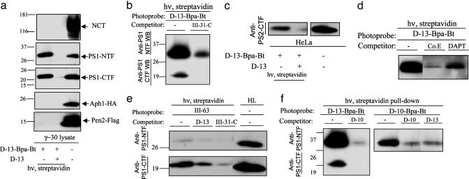
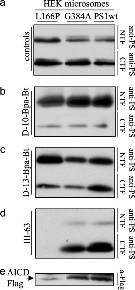
gamma-Secretase is a structurally enigmatic multiprotein complex that catalyzes intramembrane proteolysis of a variety of substrates, including the amyloid beta-protein precursor of Alzheimer's disease and the Notch receptor essential to cell differentiation. The active site of this transmembrane aspartyl protease apparently lies at the interface between two subunits of presenilin-1 (PS1); however, evidence suggests the existence of an initial substrate-binding site that is distinct from the active site. Here, we report that photoaffinity probes based on potent helical peptide inhibitors and designed to mimic the amyloid beta-protein precursor substrate bind specifically to the PS subunit interface, at a site close to the active site. The location of the helical peptide-binding site suggests that substrate passes between the two PS1 subunits to access the active site. An aggressive Alzheimer-causing mutation in PS1 strongly reduced photolabeling by a transition-state analogue but not by helical peptides, providing biochemical evidence that the pathological effect of this PS mutation is due to alteration of the active-site topography.


Figures

Fig. 1.
Fig. 1.
Photoactivatable 10-residue helical d-peptide inhibitor of γ-secretase covalently labels PS1 heterodimer at a site distinct from the active site. (a) d-10-Bpa-Bt (500 nM) was incubated with γ-30 lysates in the absence or presence of d-10 (10 μM), and the samples were irradiated at 350 nm. Biotinylated proteins were precipitated with immobilized streptavidin and blotted for NCT, PS1-NTF, PS1-CTF, the Flag-tag of Pen-2, and the HA-tag of Aph-1. Western blotting reveals that PS1-NTF is specifically tagged, but not NCT, APH-1, or Pen-2. Small traces of the labeled PS1-CTF also were observed that were only visible upon longer exposure. (b) d-10-Bpa-Bt (500 nM) was incubated with HeLa lysates in the absence or presence of d-10 (10 μM), L-10 (10 μM), and III-31-C (5 μM). (c) d-10-Bpa-Bt (500 μM) was incubated with HeLa lysates in the absence or presence of Compound E (5 μM) and DAPT (5 μM).
Fig. 2.
Fig. 2.
Photoactivatable 13-residue helical d-peptide inhibitor of γ-secretase covalently labels PS1-NTF and -CTF, but differently from 10-residue helical photoprobe. (a) d-13-Bpa-Bt (50 nM) was incubated with γ-30 lysates in the absence or presence of d-13 (250 nM), and the samples were treated and analyzed as in Fig. 1a. (b) d-13-Bpa-Bt (50 nM) was incubated with HeLa lysates in the absence or presence of III-31-C (250 nM). (c) d-13-Bpa-Bt (50 nM) was incubated with HeLa lysates in the absence or presence of d-13 (250 nM). Immunoblotting was performed with antibodies directed against the PS2 CTF. (d) d-13-Bpa-Bt (100 nM) was incubated with HeLa lysates in the absence or presence of Compound E (2 μM) and DAPT (2 μM). (e) III-63 (50 nM) was incubated with HeLa lysates in the absence or presence of d-13 (250 nM) and III-31-C (250 nM). (f)(Left) d-13-Bpa-Bt (100 nM) was incubated with HeLa lysates in the absence or presence of d-10 (2 μM). (Right) d-10-Bpa-Bt (500 nM) was incubated with HeLa lysates in the absence or presence of d-13 (5 μM) and d-10 (10 μM).
Fig. 3.
Fig. 3.
FAD-PS1 mutation L166P affects the photolabeling by the active-site-directed III-63, with little or no effect on both helical peptide photoprobes. (a) CHAPSO-solubilized membranes derived from HEK cells stably expressing PS1-L166P, PS1-G384A, and PS1-wt were normalized by the amount of PS1. (b–d) Membranes were incubated with d-10-Bpa-Bt (500 nM) (b), d-13-Bpa-Bt (500 nM) (c), and III-63 (500 nM) (d). Samples were treated and analyzed as in Fig. 1a. (e) γ-Secretase activity measured in the solubilized membranes. C100-Flag was incubated with membranes for 4 h, and APP intracellular domain (AICD)-Flag (shown), the product of the proteolysis, was detected with anti-Flag antibody.
Fig. 4.
Fig. 4.
Proposed models for inhibitors (a) and substrate (b) interactions with γ-secretase. (a)(Top) Substrate-based d-10 helical peptide interacts with PS1 at the initial binding site. (Middle) Transition-state analogue III-31-C binds to PS1 NTF/CTF heterodimer at the active site, located internally and containing two aspartates (denoted as “D”). (Bottom) d-13 peptide interacts with both active and initial binding sites. (b) Upon docking to the initial binding site, a substrate passes through PS1 subunits fully (path A) or partially (path B) to access the nearby active site.

References

    1. Esler, W. P. & Wolfe, M. S. (2001) Science 293, 1449-1454. - PubMed
    1. De Strooper, B. (2003) Neuron 38, 9-12. - PubMed
    1. Wolfe, M. S. & Kopan, R. (2004) Science 305, 1119-1123. - PubMed
    1. Yu, G., Nishimura, M., Arawaka, S., Levitan, D., Zhang, L., Tandon, A., Song, Y. Q., Rogaeva, E., Chen, F., Kawarai, T., et al. (2000) Nature 407, 48-54. - PubMed
    1. Goutte, C., Tsunozaki, M., Hale, V. A. & Priess, J. R. (2002) Proc. Natl. Acad. Sci. USA 99, 775-779. - PMC - PubMed

Publication types

LinkOut - more resources