. 2005 Mar;14(3):735-42.
doi: 10.1110/ps.041139505.

Microsecond timescale backbone conformational dynamics in ubiquitin studied with NMR R1rho relaxation experiments

Affiliations

Microsecond timescale backbone conformational dynamics in ubiquitin studied with NMR R1rho relaxation experiments

Francesca Massi et al. Protein Sci. 2005 Mar.

Abstract

NMR spin relaxation experiments are used to characterize the dynamics of the backbone of ubiquitin. Chemical exchange processes affecting residues Ile 23, Asn 25, Thr 55, and Val 70 are characterized using on- and off-resonance rotating-frame 15N R1rho relaxation experiments to have a kinetic exchange rate constant of 25,000 sec(-1) at 280 K. The exchange process affecting residues 23, 25, and 55 appears to result from disruption of N-cap hydrogen bonds of the alpha-helix and possibly from repacking of the side chain of Ile 23. Chemical exchange processes affecting other residues on the surface of ubiquitin are identified using 1H-15N multiple quantum relaxation experiments. These residues are located near or at the regions known to interact with various enzymes of the ubiquitin-dependent protein degradation pathway.


Figures

Figure 1.
Figure 1.
Conformational exchange contribution to transverse relaxation, Rex, for ubiquitin at T = 298 K (open circles) and T = 280 K (filled circles) at B0 = 11.7 T.
Figure 2.
Figure 2.
Conformational exchange contribution to transverse relaxation, Rex as a function of the effective field, ωe, at a static magnetic field of 11.7 T and T = 298 K for Ile 23 (circles) and Asn 25 (squares) in ubiquitin.
Figure 3.
Figure 3.
Cartoon representation of ubiquitin that highlights the positions of the exchanging residues Ile 23, Asn 25, Thr 55, Val 70 (A). Regions characterized by different secondary structure are represented with different colors: helices ,red; β-strands, yellow; coil and turns, blue. The boxed region is shown as an expansion in B. Each residue is labeled at the backbone N atom. The dashed lines indicate hydrogen bonds.
Figure 4.
Figure 4.
Conformational exchange contribution to transverse relaxation, Rex, as a function of the effective field, ωe at T = 280 K. Circles and squares represent data collected at a static magnetic field of 11.7 T and 14.1 T, respectively. Lines are the results of simultaneously fitting the data at 11.7 T (circles) and 14.1 T (dashed), using equation 2.
Figure 5.
Figure 5.
Amide 1H-15N differential relaxation rate constants for ZQ and DQ coherences, ΔRMQ, for ubiquitin measured at 11.7 T and T = 280 K.
Figure 6.
Figure 6.
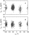
Dependence of the 15N chemical shift on side chain χ1 dihedral angle. 15N secondary chemical shifts are plotted as a function of side chain χ1 torsional angle for all isoleucine (A) and asparagine (B) residues in the RefDB chemical shift database (Zhang et al. 2003).
Figure 7.
Figure 7.
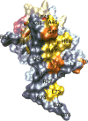
Amide 1H-15N ΔRMQ for ubiquitin measured at 11.7 T and T = 280 K. Data are represented with different colors on the surface of the protein. Residues with ΔRMQ < 3 are represented as gray, residues with 3 ≤ ΔRMQ < 5 are yellow, residues with 5 ≤ ΔRMQ < 8 are orange, residues with 8 ≤ ΔRMQ < 15 are red, and residues with ΔRMQ ≥ 15 are purple. The opaque residues are those known to be directly involved in the interaction with E1 and E2 enzymes. All other residues are depicted as transparent.

References

    1. Abragam, A. 1983. Principles of nuclear magnetism. Oxford University Press, Oxford.
    1. Beal, R., Deveraux, Q., Xia, G., Rechsteiner, M., and Pickart, C. 1996. Surface hydrophobic residues of multiubiquitin chains essential for proteolytic targeting. Proc. Natl. Acad. Sci. 93 861–866. - PMC - PubMed
    1. Braun, D., Wider, G., and Wüthrich, K. 1994. Sequence corrected 15N random coil chemical-shifts. J. Am. Chem. Soc. 116 8466–8469.
    1. Burch, T.J. and Haas, A.L. 1994. Site-directed mutagenesis of ubiquitin. Differential roles of arginine in the interaction with ubiquitin-activating enzyme. Biochemistry 33 7300–7308. - PubMed
    1. Carlomagno, T., Maurer, M., Hennig, M., and Griesinger, C. 2000. Ubiquitin backbone motion studied via NHN-C′ Cα dipolar-dipolar and C′-C′ C′/ NHN CSA-dipolar cross-correlated relaxation. J. Am. Chem. Soc. 122 5105–5113.

Publication types

MeSH terms

LinkOut - more resources