

Role of histone deacetylation in cell-specific expression of endothelial nitric-oxide synthase
- PMID: 15722551
- PMCID: PMC1283144
- DOI: 10.1074/jbc.M412960200
Role of histone deacetylation in cell-specific expression of endothelial nitric-oxide synthase
Abstract
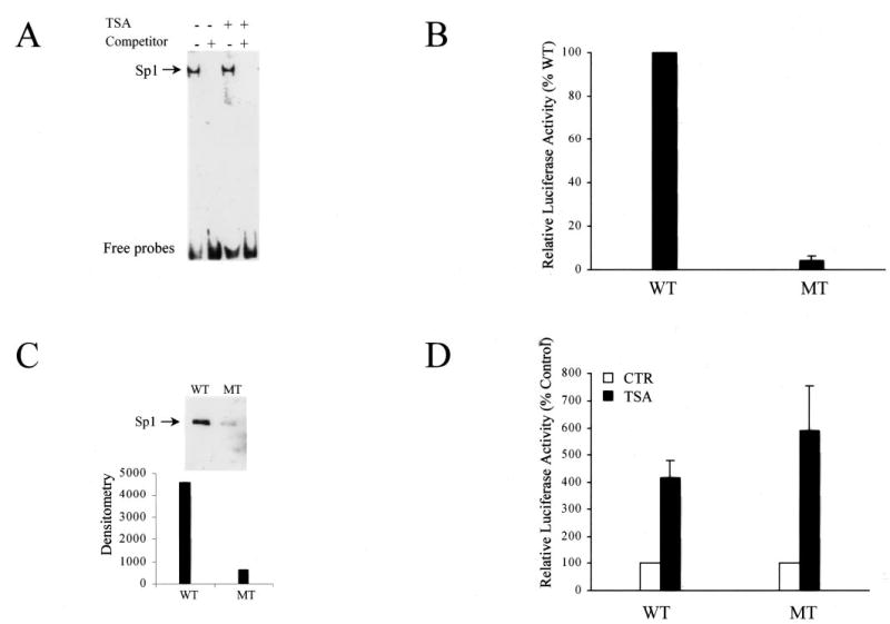
Histone acetylation plays an important role in chromatin remodeling and gene expression. The molecular mechanisms involved in cell-specific expression of endothelial nitric-oxide synthase (eNOS) are not fully understood. In this study we investigated whether histone deacetylation was involved in repression of eNOS expression in non-endothelial cells. Induction of eNOS expression by histone deacetylase (HDAC) inhibitors trichostatin A (TSA) and sodium butyrate was observed in all four different types of non-endothelial cells examined. Chromatin immunoprecipitation assays showed that the induction of eNOS expression by TSA was accompanied by a remarkable increase of acetylation of histone H3 associated with the eNOS 5'-flanking region in the non-endothelial cells. Moreover, DNA methylation-mediated repression of eNOS promoter activity was partially reversed by TSA treatment, and combined treatment of TSA and 5-aza-2'-deoxycytidine (AzadC) synergistically induced eNOS expression in non-endothelial cells. The proximal Sp1 site is critical for basal activity of eNOS promoter. The induction of eNOS by inhibition of HDACs in non-endothelial cells, however, appeared not mediated by the changes in Sp1 DNA binding activity. We further showed that Sp1 bound to the endogenous eNOS promoter and associated with HDAC1 in non-endothelial HeLa cells. Combined TSA and AzadC treatment increased Sp1 binding to the endogenous eNOS promoter but decreased the association between HDAC1 and Sp1 in HeLa cells. Our data suggest that HDAC1 plays a critical role in eNOS repression, and the proximal Sp1 site may serve a key target for HDCA1-mediated eNOS repression in non-endothelial cells.
Figures


Similar articles
-
The expression of endothelial nitric-oxide synthase is controlled by a cell-specific histone code.J Biol Chem. 2005 Jul 1;280(26):24824-38. doi: 10.1074/jbc.M502115200. Epub 2005 May 3. J Biol Chem. 2005. PMID: 15870070
-
Histone deacetylase inhibitors stimulate mitochondrial HMG-CoA synthase gene expression via a promoter proximal Sp1 site.Nucleic Acids Res. 2003 Mar 15;31(6):1693-703. doi: 10.1093/nar/gkg262. Nucleic Acids Res. 2003. PMID: 12626711 Free PMC article.
-
Inhibitors of histone deacetylation downregulate the expression of endothelial nitric oxide synthase and compromise endothelial cell function in vasorelaxation and angiogenesis.Circ Res. 2002 Nov 1;91(9):837-44. doi: 10.1161/01.res.0000037983.07158.b1. Circ Res. 2002. PMID: 12411399
-
HDAC1: an environmental sensor regulating endothelial function.Cardiovasc Res. 2022 Jun 29;118(8):1885-1903. doi: 10.1093/cvr/cvab198. Cardiovasc Res. 2022. PMID: 34264338 Free PMC article. Review.
-
Contribution of Histone Deacetylases in Prognosis and Therapeutic Management of Cholangiocarcinoma.Mol Diagn Ther. 2020 Apr;24(2):175-184. doi: 10.1007/s40291-020-00454-x. Mol Diagn Ther. 2020. PMID: 32125662 Review.
Cited by
-
Epigenetic Signatures in Arterial Hypertension: Focus on the Microvasculature.Int J Mol Sci. 2023 Mar 2;24(5):4854. doi: 10.3390/ijms24054854. Int J Mol Sci. 2023. PMID: 36902291 Free PMC article. Review.
-
Loss of methyl-CpG-binding domain protein 2 enhances endothelial angiogenesis and protects mice against hind-limb ischemic injury.Circulation. 2011 Jun 28;123(25):2964-74. doi: 10.1161/CIRCULATIONAHA.110.966408. Epub 2011 Jun 13. Circulation. 2011. PMID: 21670230 Free PMC article.
-
Endothelial heterogeneity in the umbilico-placental unit: DNA methylation as an innuendo of epigenetic diversity.Front Pharmacol. 2014 Mar 27;5:49. doi: 10.3389/fphar.2014.00049. eCollection 2014. Front Pharmacol. 2014. PMID: 24723887 Free PMC article. Review.
-
Epigenetic DNA Methylation and Protein Homocysteinylation: Key Players in Hypertensive Renovascular Damage.Int J Mol Sci. 2024 Oct 29;25(21):11599. doi: 10.3390/ijms252111599. Int J Mol Sci. 2024. PMID: 39519150 Free PMC article. Review.
-
Role of DNA methyltransferase 1 on the altered eNOS expression in human umbilical endothelium from intrauterine growth restricted fetuses.Epigenetics. 2013 Sep;8(9):944-52. doi: 10.4161/epi.25579. Epub 2013 Jul 18. Epigenetics. 2013. PMID: 23867713 Free PMC article.
References
-
- Li H, Wallerath T, Munzel T, Forstermann U. Nitric Oxide. 2002;7:149–164. - PubMed
-
- Teichert AM, Miller TL, Tai SC, Wang Y, Bei X, Robb GB, Phillips MJ, Marsden PA. Am J Physiol Heart Circ Physiol. 2000;278:1352–1361. - PubMed
-
- Guillot PV, Liu L, Kuivenhoven JA, Guan J, Rosenberg RD, Aird WC. Physiol Genomics. 2000;2:77–83. - PubMed
-
- Chan Y, Fish JE, D’Abreo C, Lin S, Robb GB, Teichert AM, Karantzoulis-Fegaras F, Keightley A, Steer BM, Marsden PA. J Biol Chem. 2004;279:35087–35100. - PubMed
Publication types
MeSH terms
Substances
Grants and funding
LinkOut - more resources
Full Text Sources
Research Materials
Miscellaneous
