

CT evaluation of the pattern of odontoid fractures in the elderly--relationship to upper cervical spine osteoarthritis
- PMID: 15723251
- PMCID: PMC3476682
- DOI: 10.1007/s00586-004-0743-z
CT evaluation of the pattern of odontoid fractures in the elderly--relationship to upper cervical spine osteoarthritis
Abstract
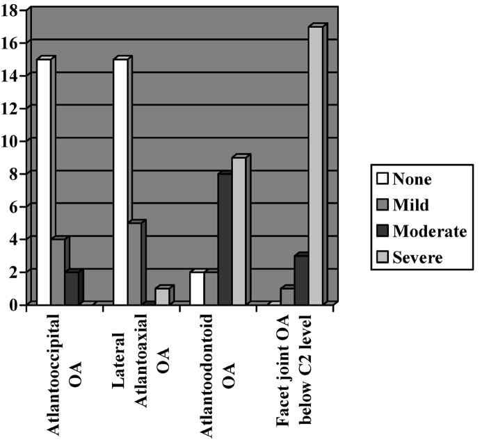
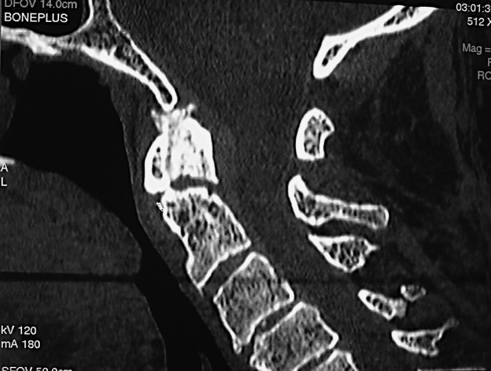
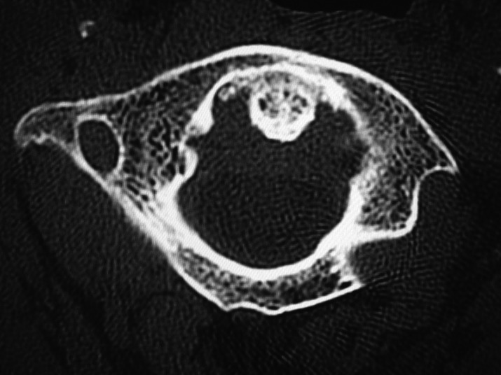
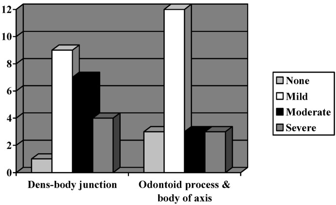
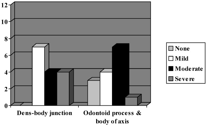
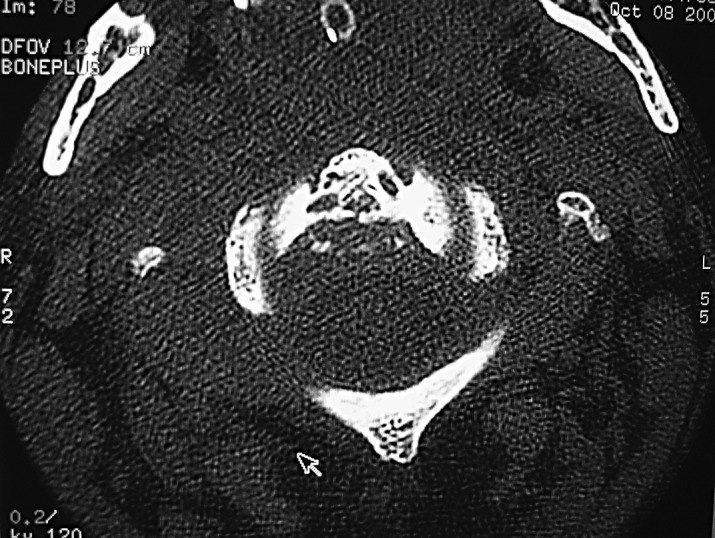
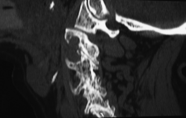
Odontoid fractures are common in the elderly following minor falls. Almost all of them have osteoarthritis of the cervical spine below the axis vertebra. As a result, there is increased stress on the spared upper cervical spine, resulting in a higher incidence of injuries. As movement in the upper cervical spine involves participation of five joints, degeneration in any one particular joint may affect the biomechanics of loading of the upper cervical spine. We aimed to analyse the relationship of odontoid fractures to the pattern of upper cervical spine osteoarthritis in the elderly. We studied the CT-scan images of the cervical spine in 23 patients who were over the age of 70 years and had odontoid fractures. In each patient, the type of odontoid fracture and the characteristics of the degenerative changes in each joint were analysed. Twenty-one of 23 patients had Type-II odontoid fractures. The incidence of significant atlanto-odontoid degeneration in these individuals was very high (90.48%), with relative sparing of the lateral atlantoaxial joints. Osteoporosis was found in 13 of 23 patients at the dens-body junction and in seven of 23 patients at the odontoid process and body of the axis. With ageing, progressively more severe degenerative changes develop in the atlanto-odontoid joint. These eventually obliterate the joint space and fix the odontoid to the anterior arch of the atlas. In contrast, the lateral atlantoaxial joints are hardly affected by osteoarthritis. Thus, ultimately, atlantoaxial movements including atlantoaxial rotation are markedly limited by osteoarthritis of the atlanto-odontoid joint. However, there is still potential for movement in the lateral atlantoaxial joints, as they remain relatively free of degenerative change. The vulnerability of the atlantoaxial segment is further increased by markedly limited rotation below the axis vertebra due to severe facet-joint degeneration. As a consequence, a relatively low-energy trauma to the lateral part of the face, for instance by a fall, will induce forced atlantoaxial rotation. This, with the marked limitation of movement at the atlanto-odontoid joint, will produce a torque force at the base of the odontoid process leading to a Type II fracture.
Figures

References
MeSH terms
LinkOut - more resources
Full Text Sources
Medical
Research Materials
