. 2005 Feb;96(2):83-92.
doi: 10.1111/j.1349-7006.2005.00012.x.

Phytosphingosine induced mitochondria-involved apoptosis

Affiliations

Phytosphingosine induced mitochondria-involved apoptosis

Yukitoshi Nagahara et al. Cancer Sci. 2005 Feb.

Abstract

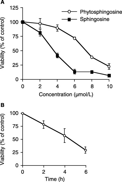
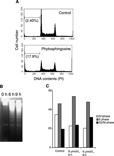
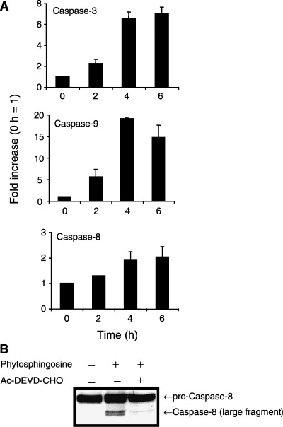
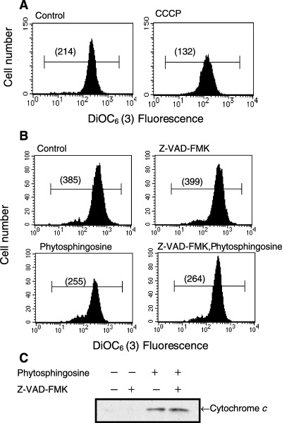
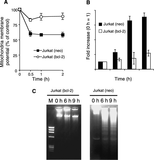
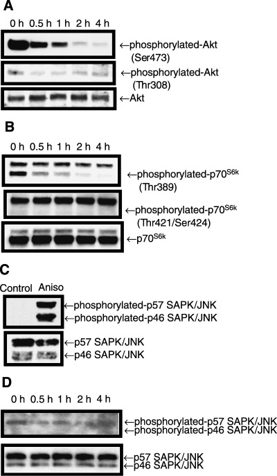
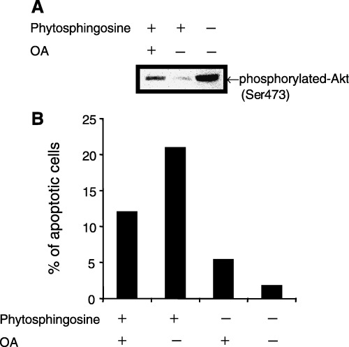
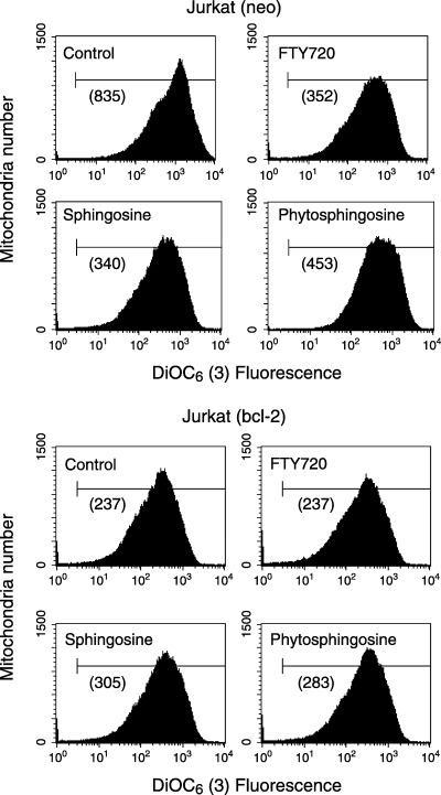
Sphingolipids are putative intracellular signal mediators in cell differentiation, growth inhibition, and apoptosis. Sphingosine, sphinganine, and phytosphingosine are structural analogs of sphingolipids and are classified as long-chain sphingoid bases. Sphingosine and sphinganine are known to play important roles in apoptosis. In the present study, we examined the phytosphingosine-induced apoptosis mechanism, focusing on mitochondria in human T-cell lymphoma Jurkat cells. Phytosphingosine significantly induced chromatin DNA fragmentation, which is a hallmark of apoptosis. Enzymatic activity measurements of caspases revealed that caspase-3 and caspase-9 are activated in phytosphingosine-induced apoptosis, but there is little activation of caspase-8 suggesting that phytosphingosine influences mitochondrial functions. In agreement with this hypothesis, a decrease in DeltaPsi(m) and the release of cytochrome c to the cytosol were observed upon phytosphingosine treatment. Furthermore, overexpression of mitochondria-localized anti-apoptotic protein Bcl-2 prevented phytosphingosine apoptotic stimuli. Western blot assays revealed that phytosphingosine decreases phosphorylated Akt and p70S6k. Dephosphorylation of Akt was partially inhibited by protein phosphatase inhibitor OA and OA attenuated phytosphingosine-induced apoptosis. Moreover, using a cell-free system, phytosphingosine directly reduced DeltaPsi(m). These results indicate that phytosphingosine perturbs mitochondria both directly and indirectly to induce apoptosis.


Figures

Figure 1
Figure 1
Dose‐ and time‐ dependent Jurkat cell viability decreases after treatment with phytosphingosine. Jurkat (neo) cells were incubated for 6 h, with or without drugs, at concentrations ranging from 2 to 10 µM (a), or for the indicated times with or without 8 µM phytosphingosine (b). At the indicated times, the cells were collected and subjected to MTT assay. The results are presented as the percentage of absorbance in untreated cultures. Each bar denotes the standard deviation (n = 4).
Figure 2
Figure 2
Phytosphingosine‐induced apoptosis and cell cycle arrest in Jurkat cells. (a) Jurkat (neo) cells were incubated with 0 or 8 µM phytosphingosine for 6 h. At the indicated times, the cells were collected and permeabilized. Cells were then stained with PI, and the DNA contents were determined by flow cytometry. Values in parentheses indicate sub‐G1 cell percentages. Data are representative of three independent experiments. (b) Jurkat (neo) cells were incubated with 8 µM phytosphingosine for 0, 6, and 9 h. At the times indicated, the cells were lyzed and DNA was prepared. DNA fragmentation was analyzed by agarose gel electrophoresis. (c) Jurkat (neo) cells were incubated with or without phytosphingosine for indicated times and doses. Percentage of cells in each cell cycle phase were measured. Data are representative of three independent experiments.
Figure 3
Figure 3
Phytosphingosine‐induced caspase activation. (a) Jurkat (neo) cells were incubated with 8 µM phytosphingosine for 0, 2, 4, and 6 h. At the times indicated, cells were lyzed and caspase‐3, caspase‐9, and caspase‐8 activity were determined by specific peptides, Ac‐DEVD‐MCA, Ac‐LEHD‐MCA, and Ac‐IETD‐MCA, respectively. Each bar denotes the standard deviation (n = 3). (b) Jurkat (neo) cells were incubated with or without 20 µM Ac‐DEVD‐CHO for 1 h, thereafter with or without 8 µM phytosphingosine for 4 h. Cells were collected and lyzed. Western blots were performed with antibody for caspase‐8.
Figure 4
Figure 4
Phytosphingosine affected the mitochondria independent of caspases. (a) Jurkat (neo) cells were incubated with or without 5 µM CCCP for 30 min. DiOC6 (3) was added 15 min before the end of the culture period, and ΔΨm was analyzed by flow cytometry. Values in parentheses indicate the median fluorescence intensity. Data are representative of three independent experiments. (b) Jurkat (neo) cells were incubated with or without 50 µM Z‐VAD‐FMK for 30 min, thereafter with or without 8 µM phytosphingosine for 30 min. DiOC6 (3) was added 15 min before the end of the culture period, and ΔΨm was analyzed by flow cytometry. Values in parentheses indicate the median fluorescence intensity. Data are representative of three independent experiments. (c) Jurkat (neo) cells were incubated with or without 50 µM Z‐VAD‐FMK for 30 min, thereafter with or without 8 µM phytosphingosine for 4 h. Cells were collected and cytosol fraction was obtained. Western blots were performed with antibody for cytochrome c.
Figure 5
Figure 5
Phytosphingosine‐induced apoptosis is blocked by overexpression of Bcl‐2. (a) Jurkat (neo) and Jurkat (bcl‐2) cells were incubated with 8 µM phytosphingosine for 0, 0.5, 1, and 2 h. DiOC6 (3) was added 15 min before the end of the culture period, and ΔΨm was analyzed by flow cytometry. The results are presented as averages of the median fluorescence intensity. Each bar denotes the standard deviation (n = 3). (b) Jurkat (neo) and Jurkat (bcl‐2) cells were incubated with 8 µM phytosphingosine for 0, 2, 4, and 6 h. At the times indicated, cells were lyzed and caspase‐3 activity was determined by specific peptide, Ac‐DEVD‐MCA. Each bar denotes the standard deviation (n = 3). (c) Jurkat (neo) and Jurkat (bcl‐2) cells were incubated with 8 µM phytosphingosine for 0, 6, and 9 h. At the times indicated, the cells were lyzed and DNA was prepared. DNA fragmentation was analyzed by agarose gel electrophoresis.
Figure 6
Figure 6
Phytosphingosine reduced phosphorylated Akt and p70S6k. Jurkat (neo) cells were incubated with 8 µM phytosphingosine for the indicated times. Cells were collected and lyzed. Western blots were performed with antibodies specific for phosphorylated Akt (Ser473) (Thr308) and Akt (a), phosphorylated p70S6k (Thr389) (Thr421/Ser424), and p70S6k (b), and phosphorylated SAPK/JNK (Thr183/Tyr185) and SAPK/JNK (d). For positive control of phosphorylated SAPK/JNK (Thr183/Thr185), 1 h incubation with 1 µg/mL anisomysin (Aniso) was used (c). Samples were exposed to films for 30 s, except when detecting phosphorylated SAPK/JNK in (d), which was exposed to film for 5 min.
Figure 7
Figure 7
OA attenuated phytosphingosine‐induced Akt phosphorylation and apoptosis. (a) Jurkat (neo) cells were incubated with 400 nM OA for 30 min, thereafter 8 µM phytosphingosine for 1 h. Cells were collected and lyzed. Western blots were performed with antibodies specific for phosphorylated Akt (Ser473). (b) Jurkat (neo) cells were incubated with 400 nM OA for 30 min, thereafter 8 µM phytosphingosine for 4 h. At the indicated times, cells were collected and stained with Hoechst 33342 and PI. Cells were counted using fluorescence microscopy and percentage of blue‐ and red‐stained bubbled nuclei cells (apoptotic cells) in whole cells were estimated. Data are representative of three independent experiments.
Figure 8
Figure 8
Phytosphingosine reduced ΔΨm directly in isolated Jurkat mitochondria. Mitochondria from Jurkat (neo) and (bcl‐2) cells (25 µg mitochondrial protein in 100 µL CFS buffer) were prepared as described in Materials and Methods. Mitochondria were incubated with no additives, 10 µM phytosphingosine, 10 µM sphingosine, and 10 µM FTY720 (positive control) for 30 min at 37°C. Mixtures were then incubated with DiOC6(3) for 15 min, and ΔΨm was analyzed by flow cytometry. Values in parentheses indicate the median fluorescence intensity. (a), Jurkat (neo) mitochondria; (B), Jurkat (bcl‐2) mitochondria. Data are representative of two independent experiments.

References

    1. Zamzami N, Marzo I, Susin SA, Brenner C, Larochette N, Marchetti P, Reed J, Kofler R, Kroemer G. The thiol crosslinking agent diamide overcomes the apoptosis‐inhibitory effect of Bcl‐2 by enforcing mitochondrial permeability transition. Oncogene 1998; 16: 1055–63. - PubMed
    1. Narita M, Shimizu S, Ito T, Chittenden T, Lutz RJ, Matsuda H, Tsujimoto Y. Bax interacts with the permeability transition pore to induce permeability transition and cytochrome c release in isolated mitochondria. Proceedings Natl Acad Sci USA 1998; 95: 14681–6. - PMC - PubMed
    1. Liu X, Kim CN, Yang J, Jemmerson R, Wang X. Induction of apoptotic program in cell‐free extracts: requirement for dATP and cytochrome c . Cell 1996; 86: 147–57. - PubMed
    1. Susin SA, Lorenzo HK, Zamzami N, Marzo I, Snow BE, Brothers GM, Mangion J, Jacotot E, Costantini P, Loeffler M, Larochette N, Goodlett DR, Aebersold R, Siderovski DP, Penninger JM, Kroemer G. Molecular characterization of mitochondrial apoptosis‐inducing factor. Nature 1999; 397: 441–6. - PubMed
    1. Verhagen AM, Ekert PG, Pakusch M, Silke J, Connolly LM, Reid GE, Moritz RL, Simpson RJ, Vaux DL. Identification of DIABLO, a mammalian protein that promotes apoptosis by binding to and antagonizing IAP proteins. Cell 2000; 102: 43–53. - PubMed

Publication types

MeSH terms