

Ventricular structure and function in aged dogs with renal hypertension: a model of experimental diastolic heart failure
- PMID: 15723971
- PMCID: PMC1805473
- DOI: 10.1161/01.CIR.0000157183.21404.63
Ventricular structure and function in aged dogs with renal hypertension: a model of experimental diastolic heart failure
Abstract
Background: Heart failure (HF) with normal ejection fraction (diastolic HF [DHF]) usually occurs in elderly patients with hypertension. The presence and significance of altered systolic and diastolic ventricular function in DHF is increasingly controversial. Our objective was to develop a clinically relevant large-animal model to better understand the pathophysiology of DHF.
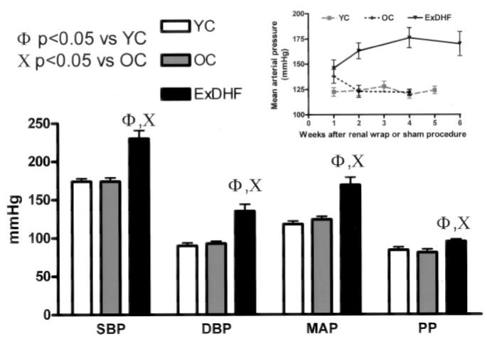
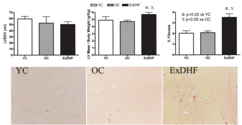
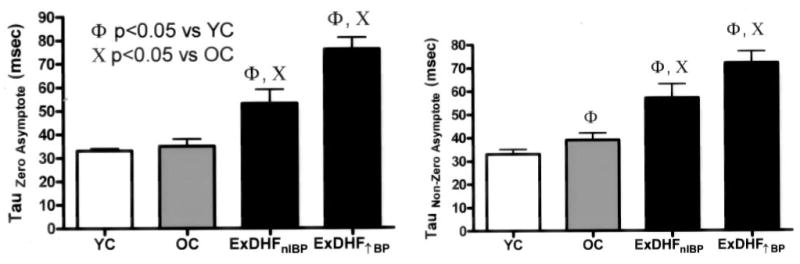
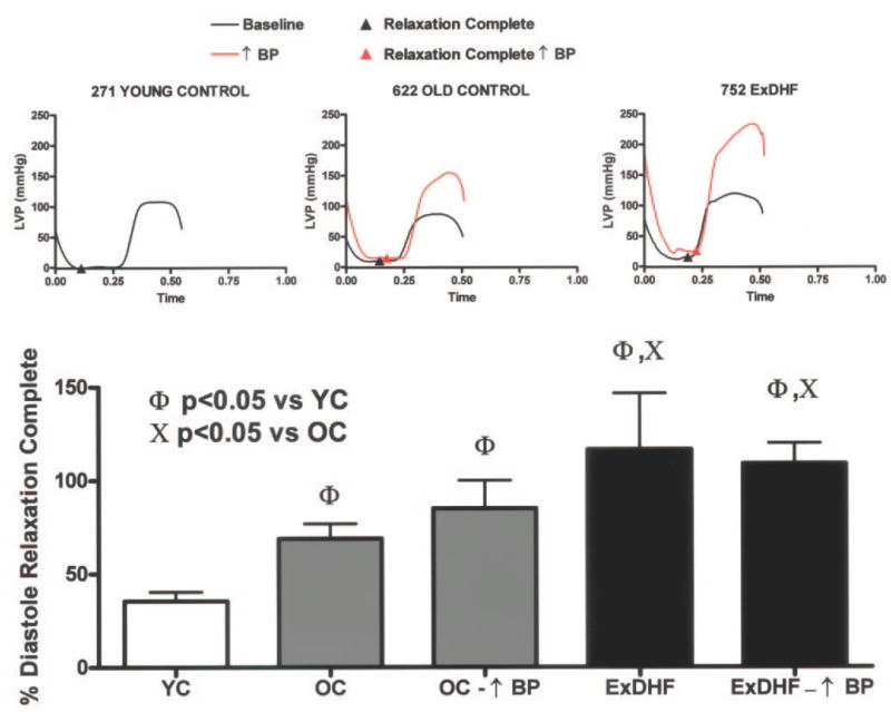
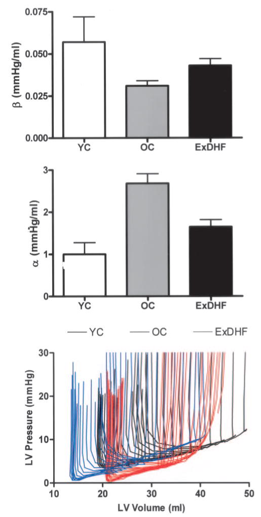
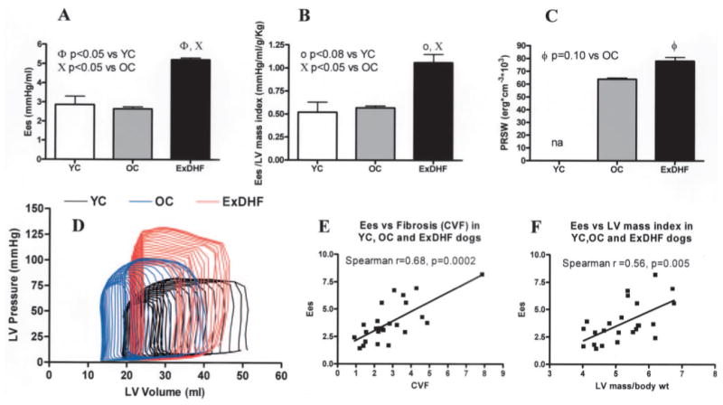
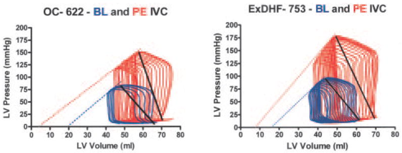
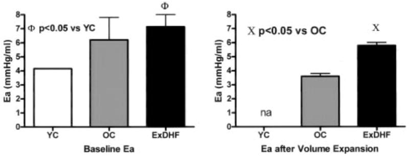
Methods and results: Ventricular structure and function were characterized in young control (YC group; n=6), old control (OC group; n=7), and old dogs made hypertensive by renal wrapping (experimental DHF [ExDHF] group; n=8). The ExDHF group was associated with normal left ventricular (LV) volume, increased LV mass, and myocardial fibrosis. LV relaxation was impaired in ExDHF (tau=53+/-6 ms) compared with OC (tau=35+/-3 ms; P<0.05) and YC (tau=33+/-6 ms; P<0.05) dogs. The percent diastole at which relaxation is complete was increased in ExDHF (116+/-30%) compared with OC (69+/-8%; P<0.05) and YC (35+/-5%; P<0.05) dogs. The coefficient of LV diastolic stiffness was similar in OC, YC, and ExDHF dogs. Diastolic pressures increased dramatically in response to increases in blood pressure. End-systolic LV stiffness was enhanced in ExDHF dogs and after load enhancement of myocardial performance was maintained. Arterial stiffness was increased in ExDHF dogs.
Conclusions: Aged dogs with chronic hypertension exhibit LV hypertrophy and fibrosis with impaired LV relaxation but no increase in the coefficient of LV diastolic stiffness. LV systolic and arterial stiffness are increased, which may exacerbate load-dependent impairment of relaxation and contribute to increased filling pressures with hypertensive episodes. This model mimics many of the structural and functional characteristics described in the limited studies of human DHF and provides insight into the pathogenesis of DHF.
Figures
References
-
- Hart CY, Redfield MM. Diastolic heart failure in the community. Current Cardiol Reports. 2000;2:461–469. - PubMed
-
- Zile MR, Brutsaert DL. New concepts in diastolic dysfunction and diastolic heart failure, part II: causal mechanisms and treatment. Circulation. 2002;105:1503–1508. - PubMed
-
- Zile MR, Gaasch WH, Carroll JD, Feldman MD, Aurigemma GP, Schaer GL, Ghali JK, Liebson PR. Heart failure with normal ejection fraction: is measurement of diastolic function necessary to make the diagnosis of diastolic heart failure? Circulation. 2001;104:779–782. - PubMed
-
- Zile MR, Baicu CF, Gaasch WH. Diastolic heart failure: abnormalities in active relaxation and passive stiffness of the left ventricle. N Engl J Med. 2004;350:1953–1959. - PubMed
-
- Kawaguchi M, Hay I, Fetics B, Kass DA. Combined ventricular systolic and arterial stiffening in patients with heart failure and preserved ejection fraction. Circulation. 2003;107:714–720. - PubMed
Publication types
MeSH terms
Grants and funding
LinkOut - more resources
Full Text Sources
Medical
Research Materials
Miscellaneous
