. 2005 Jul 1;389(Pt 1):27-35.
doi: 10.1042/BJ20050223.

Identification and functional characterization of a novel Candida albicans gene CaMNN5 that suppresses the iron-dependent growth defect of Saccharomyces cerevisiae aft1Delta mutant

Affiliations

Identification and functional characterization of a novel Candida albicans gene CaMNN5 that suppresses the iron-dependent growth defect of Saccharomyces cerevisiae aft1Delta mutant

Chen Bai et al. Biochem J. .

Abstract

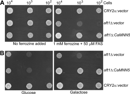
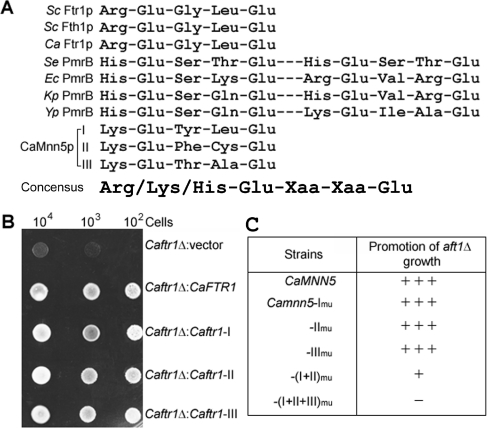
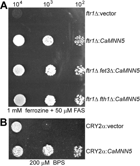
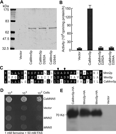
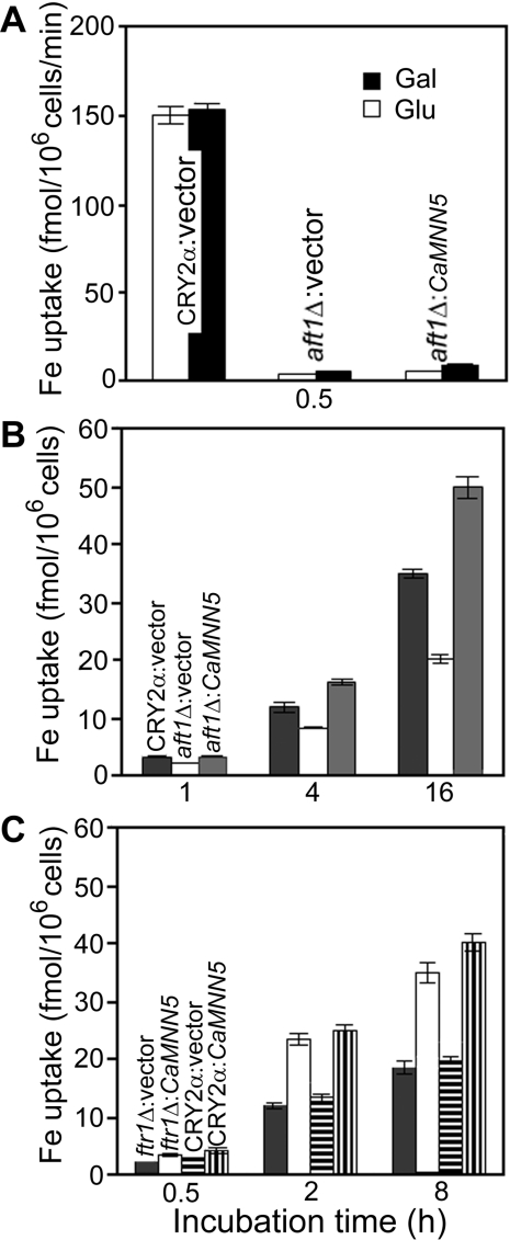
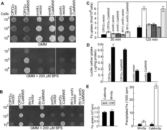
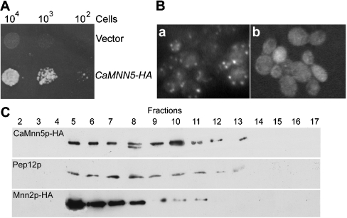
In Saccharomyces cerevisiae, the transcription factor Aft1p plays a central role in regulating many genes involved in iron acquisition and utilization. An aft1Delta mutant exhibits severely retarded growth under iron starvation. To identify the functional counterpart of AFT1 in Candida albicans, we transformed a C. albicans genomic DNA library into aft1Delta to isolate genes that could allow the mutant to grow under iron-limiting conditions. In the present paper, we describe the unexpected discovery in this screen of CaMNN5. CaMnn5p is an alpha-1,2-mannosyltransferease, but its growth-promoting function in iron-limiting conditions does not require this enzymatic activity. Its function is also independent of the high-affinity iron transport systems that are mediated by Ftr1p and Fth1p. We obtained evidence suggesting that CaMnn5p may function along the endocytic pathway, because it cannot promote the growth of end4Delta and vps4Delta mutants, where the endocytic pathway is blocked at an early and late step respectively. Neither can it promote the growth of a fth1Delta smf3Delta mutant, where the vacuole-cytosol iron transport is blocked. Expression of CaMNN5 in S. cerevisiae specifically enhances an endocytosis-dependent mechanism of iron uptake without increasing the uptake of Lucifer Yellow, a marker for fluid-phase endocytosis. CaMnn5p contains three putative Lys-Glu-Xaa-Xaa-Glu iron-binding sites and co-immunoprecipitates with 55Fe. We propose that CaMnn5p promotes iron uptake and usage along the endocytosis pathway under iron-limiting conditions, a novel function that might have evolved in C. albicans.


Figures

Figure 1
Figure 1. CaMNN5 enhances aft1Δ growth on iron-limiting media
(A) CaMNN5 on a 2μ vector with its native promoter (aft1Δ:CaMNN5) suppressed the growth defect of aft1Δ on the iron-limiting GMM plate containing 1 mM ferrozine and 50 μM ferrous ammonium sulphate (FAS). The wild-type strain and aft1Δ transformed with the empty vector (CRY2α:vector and aft1Δ:vector respectively) were included for comparison. The GMM plate without ferrozine is iron-sufficient. (B) aft1Δ transformed with a CEN plasmid harbouring CaMNN5 driven by the Gal1-10 promoter (aft1Δ:CaMNN5) grew on the iron-limiting GaMM, but not on GMM plates. CRY2α and aft1Δ transformed with the empty plasmid were used as controls. Approx. 104, 103 and 102 cells of each strain were spotted on to the plates and incubated at 30 °C for 5 days.
Figure 2
Figure 2. CaMnn5p contains three potential iron-binding Lys-Glu-Xaa-Xaa-Glu motifs
(A) The Lys-Glu-Xaa-Xaa-Glu motifs of CaMnn5p were aligned with some conserved motifs of several known iron-binding proteins: S. cerevisiae (Sc) Ftr1p (amino acids 157–161) and Fth1p (amino acids 242–246), C. albicans (Ca) Ftr1p (amino acids 157–161), Salmonella enterica (Se) PmrB (amino acids 38–42, 63–67), Escherichia coli (Ec) PmrB (amino acids 38–42, 60–64), Klebsiella pneumoniae (Kp) PmrB (amino acids 36–40, 62–66) and Yersinia pestis (Yp) PmrB (amino acids 29–33, 54–58). (B) Each of the Lys-Glu-Xaa-Xaa-Glu motifs of CaMnn5p could functionally replace the Arg157-Glu-Gly-Leu-Glu161 motif of CaFtr1p. All strains were grown on iron-limiting GMM plates at 30 °C for 5 days. (C) The Lys-Glu-Xaa-Xaa-Glu motifs of CaMnn5p were mutated by site-directed mutagenesis. Cells of each strain were spotted on to iron-limiting GMM and GaMM plates for incubation at 30 °C for 5 days. The table summarizes the ability of each mutated gene in promoting aft1Δ growth on the GaMM plates. None of the strains grew on the GMM plates.
Figure 3
Figure 3. CaMNN5 promotes cell growth under iron-limiting conditions by a mechanism independent of the high-affinity iron transporters
(A) CaMNN5 expression promoted the growth on iron-limiting plates of S. cerevisiae mutants deleted of high-affinity iron transporter genes, such as ftr1Δ, ftr1Δ fet3Δ and ftr1Δ fth1Δ. CaMNN5 was transformed into each mutant on the CEN plasmid driven by the Gal1-10 promoter. The cells were spotted on to iron-limiting GaMM plates. (B) CaMNN5 promoted the growth of the wild-type strain under more stringent iron-limiting conditions. The same plasmid expressing CaMNN5 described in (A) was transformed into CRY2α, and the transformants were grown on GaMM containing 200 μM BPS. The plate was incubated at 30 °C for 5 days.
Figure 4
Figure 4. Mannosyltransferase activity of CaMnn5p is not required for promoting cell growth
(A) CaMnn5p and two mutant forms were expressed in P. pastoris. Aliquots of 20 μl of culture supernatant from each of the strains expressing CaMnn5p, CaMnn5p (D282A) or CaMnn5p (D284A) [see below for the description of the mutants in (C)] were examined by SDS/10% PAGE. The gel was stained with Coomassie Blue. (B) The supernatants from the same cultures described in (A) were assayed directly for mannosyltransferase activity. The assay was conducted three times for each sample and results are means±S.D. The results are expressed as activity/mg of protein per h. Protein concentration was determined by the Bradford assay. (C) Amino acid sequence alignment of a segment of the catalytic region of three α-1,2-mannosyltransferases, S. cerevisiae Mnn2p and Mnn5p and C. albicans CaMnn5p. The arrows indicate two essential aspartate residues. (D) Expression of MNN2 and MNN5 did not promote the growth of aft1Δ on the iron-limiting plates. CaMNN5, MNN2 and MNN5 were cloned on the same CEN plasmid driven by the Gal1-10 promoter. Each was transformed into aft1Δ, and the transformants were spotted on to iron-limiting GaMM plates. FAS, ferrous ammonium sulphate. (E) HA-tagged CaMNN5, MNN5 and MNN2 were transformed into aft1Δ. The transformants were grown in liquid GaMM before total protein was extracted for Western blot analysis.
Figure 5
Figure 5. CaMNN5 enhances a slow process of iron uptake
(A) Strains CRY2α:vector, aft1Δ:vector and aft1Δ:CaMNN5 driven by the Gal1-10 promoter were first grown in GaMM to exponential phase. Then the same number of cells of each strain was assayed for iron uptake in the same medium supplemented with 2 μM 59Fe for 30 min. 59Fe uptake was determined by scintillation counting and expressed as fmol/106 cells per min. (B) The same strains used in (A) were incubated in GaMM containing 200 μM BPS and 2 μM 59Fe for 1, 4 and 16 h. The iron uptake is expressed as the total amount of 59Fe accumulated in 106 cells at the end of incubation. (C) Strains CRY2α and ftr1Δ transformed with CaMNN5 driven by the Gal1-10 promoter or the empty vector were incubated in GaMM containing 200 μM BPS and 2 μM 59Fe for 0.5, 2 and 8 h. Each assay was repeated three times.
Figure 6
Figure 6. CaMnn5p-mediated iron uptake in mutants defective of the endocytic pathway
(A) CRY2α, end4Δ and vps4Δ mutants were transformed with a 2μ plasmid carrying CaMNN5 under the control of FTR1 promoter or with the empty plasmid. The cells were spotted on to GMM plates containing no chelator or 200 μM BPS and incubated at 25 °C for 7 days. (B) CRY2α, fth1Δ, smf3Δ and fth1Δ smf3Δ strains were transformed with the same plasmids described in (A) and grown on GMM plates containing 200 μM BPS at 30 °C for 5 days. (C) 59Fe uptake assay was performed in GMM containing 200 μM BPS and 2 μM 59Fe for 30 and 120 min. The total amount of 59Fe accumulated at the end of incubation is shown. The amount of 59Fe associated with the cells kept on ice was subtracted. (D) LY uptake into CRY2α or end4Δ cells transformed with CaMNN5 or vector alone. There was no detectable LY uptake when the cells were kept on ice. The levels of cellular LY are expressed as μg of LY/mg of protein. (E) Co-immunoprecipitation of 55Fe with CaMnn5p–HA and Mnn5p–HA. The left-hand panel shows the amount of cellular 55Fe accumulation as c.p.m./g of cell pellet, and the inset shows the expression levels of the two HA-tagged proteins determined by Western blot analysis. The right-hand panel shows the amount of 55Fe (c.p.m./g of cell pellet) co-immunoprecipitated with CaMnn5p–HA and Mnn5p–HA. The HA-tagged protein was precipitated by mixing cell lysate with anti-HA antibody-coated agarose beads (open bars) or with naked agarose beads (closed bars). All assays were repeated three times.
Figure 7
Figure 7. Subcellular localization of CaMnn5p
(A) CaMnn5p was tagged with HA at the C-terminal end and expressed from a CEN plasmid under the control of the Gal1-10 promoter. (B) Indirect immunofluorescence staining of the CaMnn5p–HA-expressing cells. a, anti-HA antibody was used as the primary antibody; b, the primary antibody was omitted. (C) The subcellular localization of CaMnn5p–HA was determined by using sucrose gradient fractionation and Western blot analysis. The fractionation patterns of Mnn2p and Pep12p were also determined for comparison.

Similar articles

Cited by

References

    1. Hill H. A. O. Iron: an element well-fitted for its task? In: Dunford H. B., Dolphin D., Raymond K. N., Seiker L., editors. The Biological Chemistry of Iron. Reidel, Dordrecht; 1982. pp. 1–12.
    1. Crichton R. R., Charloteaux-Wauters M. Iron transport and storage. Eur. J. Biochem. 1987;164:485–506. - PubMed
    1. Elin R. J., Wolff S. M. Effect of pH and iron concentration on growth of Candida albicans in human serum. J. Infect. Dis. 1973;127:705–708. - PubMed
    1. Ramanan N., Wang Y. A high-affinity iron permease essential for Candida albicans virulence. Science. 2000;288:1062–1064. - PubMed
    1. Griffiths E. Iron in biological systems. In: Bullen J. J., Griffiths E., editors. Iron and Infection: Molecular, Clinical, and Physiological Aspects. Chichester: Wiley; 1999. pp. 1–25.

MeSH terms