. 2005 Jul 1;389(Pt 1):99-110.
doi: 10.1042/BJ20041287.

The tetraspanin D6.1A and its molecular partners on rat carcinoma cells

Affiliations

The tetraspanin D6.1A and its molecular partners on rat carcinoma cells

Christoph Claas et al. Biochem J. .

Abstract

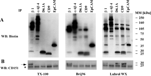
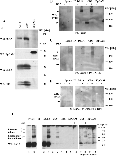
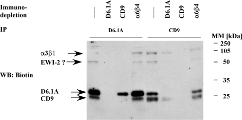
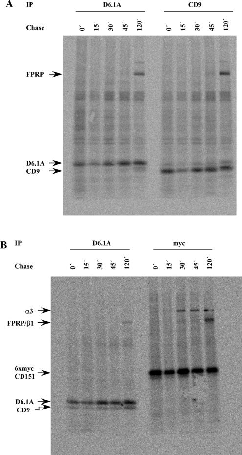
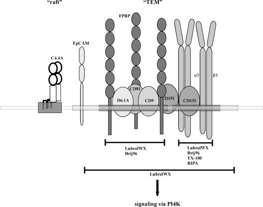
Tetraspanins function as molecular organizers of multi-protein complexes by assembling primary complexes of a relatively low mass into extensive networks involved in cellular signalling. In this paper, we summarize our studies performed on the tetraspanin D6.1A/CO-029/TM4SF3 expressed by rat carcinoma cells. Primary complexes of D6.1A are almost indistinguishable from complexes isolated with anti-CD9 antibody. Indeed, both tetraspanins directly associate with each other and with a third tetraspanin, CD81. Moreover, FPRP (prostaglandin F2alpha receptor-regulatory protein)/EWI-F/CD9P-1), an Ig superfamily member that has been described to interact with CD9 and CD81, is also a prominent element in D6.1A complexes. Primary complexes isolated with D6.1A-specific antibody are clearly different from complexes containing the tetraspanin CD151. CD151 is found to interact only with D6.1A if milder conditions, i.e. lysis with LubrolWX instead of Brij96, are applied to disrupt cellular membranes. CD151 probably mediates the interaction of D6.1A primary complexes with alpha3beta1 integrin. In addition, two other molecules were identified to be part of D6.1A complexes at this higher level of association: type II phosphatidylinositol 4-kinase and EpCAM, an epithelial marker protein overexpressed by many carcinomas. The characterization of the D6.1A core complex and additional more indirect interactions will help to elucidate the role in tumour progression and metastasis attributed to D6.1A.


Figures

Figure 1
Figure 1. D6.1A complexes resemble CD9 complexes, but are distinct from CD151 complexes
(A) PROb cells were surface-labelled with biotin and then lysed in a buffer containing 1% Triton X-100, 1% Brij96 or 1% LubrolWX. Lysates were cleared by centrifugation at 20000 g for 10 min. Equal portions of the lysates were immunoprecipitated with antibodies directed to the indicated antigens. Isolated material was separated by SDS/PAGE on a 4–20% linear gradient gel, transferred to nitrocellulose and visualized by Extravidin–HRP, followed by chemiluminescence. (B) The membrane depicted in (A) was then incubated with an antiserum recognizing the EC2 domain of rat CD151, and bands were visualized by incubation with anti-rabbit IgG–HRP. A band corresponding to CD151 is marked with an arrow; the asterisk indicates an unspecific band occurring in all lanes.
Figure 2
Figure 2. D6.1A directly associates with CD9, CD81 and FPRP
(A) PROb cells were lysed in 1% Brij96 and immunoprecipitations (IP) were performed with anti-D6.1A, anti-CD9 and anti-EpCAM antibodies. After separation of the isolated complexes by SDS/PAGE and transfer to a nitrocellulose membrane, Western blotting (WB) was performed with anti-FPRP, anti-EpCAM, anti-D6.1A and anti-CD9. (B) Brij96 lysates of PROb cells were treated with 0.5 mM of the reducible cross-linker DTSP (‘DSP’) in DMSO or with DMSO only for 1 h at room temperature, and immunoprecipitations were performed with D6.1A-, CD9- and EpCAM-specific antibodies. Samples were separated by SDS/PAGE (6% polyacrylamide), transferred to nitrocellulose and immunoblotted with anti-FPRP. In parallel, an aliquot of DTSP-treated lysate and control lysate was analysed. The arrow indicates cross-linked material containing FPRP at a mass well beyond 200 kDa; the arrowhead indicates the position of non-cross-linked FPRP at approx. 120 kDa. (C) Same as in (B), but lysates were incubated with 1% Triton X-100 after cross-linking with DTSP to remove any non-cross-linked proteins from the precipitated material. (D) Same as in (C), but samples were separated under reducing conditions (100 mM dithiothreitol) to separate the cross-linked proteins before SDS/PAGE and blotting. (E) PROb cells were lysed in 1% Brij96 and immunoprecipitations were performed with antibodies as in (AD) and, additionally, with anti-CD81 (lanes 3–10). In parallel, the lysate was analysed in the same way (lanes 1 and 2). Complexes were separated on an 11% gel and immunoblotted with anti-D6.1A. Right panel: lanes 7′, 8′, 9′ and 10′ show a longer exposure of lanes 7–10.
Figure 3
Figure 3. Stoichiometry of the association of D6.1A with CD9
PROb cells were surface-biotinylated and lysed in 1% Brij96. The lysate was divided into four equal aliquots and depleted with Protein G–Sepharose alone or with Protein G–Sepharose coupled with a monoclonal antibody specific for D6.1A, CD9 and α6β4 integrin respectively. After four successive rounds of immunodepletion, immunoprecipitations were performed with anti-D6.1A or anti-CD9 on the lysates. Complexes were separated by SDS/PAGE (12% polyacrylamide) and the biotinylated proteins were revealed by blotting with Extravidin–HRP. The question mark indicates that the identity of the 70 kDa protein as EWI-2 has yet to be verified, although work published by other groups has suggested that this protein could be EWI-2 (see the Discussion).
Figure 4
Figure 4. Analysis of D6.1A complexes by gel filtration on a Sepharose CL6B column
Brij96 lysate from approx. 2×10⁷ biotinylated PROb cells was fractioned on a Sepharose CL6B column (30 cm×0.8 cm). Fractions were collected from the leading edge of the Blue Dextran (approximately the V0) to Phenol Red (indicating the small molecule elution point). Aliquots of each fraction were immunoprecipitated with anti-D6.1A antibody and immunoblotted for biotinylated material with Extravidin–HRP. α3 and β1 integrins are found mainly in fractions V0, 1 and 2. The elution profile of total D6.1A- and D6.1A-associated FPRP was also analysed by immunoblotting with the respective antibodies.
Figure 5
Figure 5. Association of D6.1A and CD9 with FPRP during biosynthesis
PROb cells (A) or PROb cells expressing 6×mycCD151 (B) were starved for 2 h and then labelled with 0.5 mCi of [35S]Met/Cys in 1 ml of RPMI 1640 containing 5% dialysed FCS for 15 min at 37 °C (zero time=0 min, 0′). Cells were subsequently incubated in complete medium containing a 25-fold excess of unlabelled methionine and cysteine for various time periods. At the indicated time points, cells were put on ice and lysed with a buffer containing 1% Brij96. Immunoprecipitations were performed with monoclonal antibodies recognizing D6.1A, CD9 or a Myc tag attached to CD151. Samples were separated on a 5–15% linear gradient gel, transferred to nitrocellulose and exposed to a phosphoimager screen for 3 days. The radiolabelled band at 120 kDa found in D6.1A and CD9 immunoprecipitates is FPRP. This was verified by reprecipitation with FPRP-specific serum after elution of the associated protein from D6.1A and CD9 complexes using RIPA buffer (results not shown).
Figure 6
Figure 6. EpCAM is a low stoichiometric partner in D6.1A complexes
(A) Biotinylated PROb cells were lysed in 1% LubrolWX and immunodepletion was performed in four successive rounds with the indicated antibodies coupled with Protein G–Sepharose or Protein G–Sepharose alone. Equal aliquots of these lysates were subjected to immunoprecipitation with anti-D6.1A and anti-EpCAM antibodies. Precipitates were separated by SDS/PAGE (12% polyacrylamide) and then the biotinylated material was transferred to a nitrocellulose membrane and detected with Extravidin–HRP. (B) Immunoprecipitations with anti-α2 integrin, anti-α3 integrin, anti-α6β4 integrin, anti-D6.1A, anti-CD9 and anti-EpCAM antibodies were performed on 1% LubrolWX lysates of PROb cells and the biotinylated proteins were detected as described for Figure 1(A). (C) BSp73ASML cells were lysed in 1% LubrolWX (Lu) or 1% Brij98 (B'98) and immunoprecipitations were performed with anti-α6β4, anti-D6.1A, anti-CD9 and anti-EpCAM. In vitro PI4K assays were performed on the samples as described in the Materials and methods section. Phosphorylated lipids were separated by TLC and revealed by autoradiography.
Figure 7
Figure 7. Association of D6.1A and EpCAM with DRM under various detergent conditions
(AC) BSp73ASML cells were lysed in a buffer containing 1% LubrolWX (A), 1% Brij96 (B) or 1% Triton X-100 (C). Lysates were cleared by centrifugation at 20000 g for 10 min and the supernatants corresponding to approx. 2×10⁷ cells were adjusted to 40% sucrose. The sucrose-containing supernatant was layered on to a cushion of 50% sucrose and successively overlaid with a buffer containing 30, 20 and 5% sucrose respectively. Gradients thus formed were centrifuged at 217000 g for 18 h in a Kontron TST60.4 rotor. Eight fractions of equal volume were removed with a pipette from the top of each gradient and the pellet was resuspended in Laemmli sample buffer. Equal volumes of each fraction and the pellet were resolved by SDS/PAGE, transferred to nitrocellulose and immunoblotted with antibodies directed to the indicated antigens. (DF) PROb cells were surface-biotinylated and lysed in a buffer containing 1% LubrolWX (D), 1% Brij96 (E) or 1% Triton X-100 (F). Immunoprecipitations were performed with anti-D6.1A, anti-α6β4, anti-EpCAM and anti-C4.4A antibodies. Precipitates were separated on a 5–15% linear gradient gel and biotinylated proteins were revealed as described for Figure 1(A).
Figure 8
Figure 8. Analysis of D6.1A complexes isolated from LubrolWX lysates, by gel filtration on a Sepharose CL4B column
(A) Surface-biotinylated 804G cells (∼2×10⁷) were lysed in 1% LubrolWX and fractionated on a Sepharose CL4B column (50 cm×1 cm). Fractions of approx. 1 ml each were collected and void volume and the small molecule elution point were determined as described for Figure 4. Aliquots of each fraction were immunoprecipitated with anti-D6.1A and the associated biotinylated material was revealed by Extravidin–HRP. The distribution of total D6.1A, C4.4A and caveolin was determined by blotting aliquots of each fraction with the respective antibodies. (B) Fractions corresponding to the void volume (V0+1) and from within the gradient (6+7) found to contain peaks of D6.1A-associated proteins as shown in (A) were pooled and subjected to ultracentrifugation at 100000 g for 2 h in a SW41 rotor. The pellet was washed once with lysate buffer and then solubilized in 1% LubrolWX+0.1% SDS. Equal volumes of supernatant (S) and pellet (P) were precipitated with chloroform/methanol, separated on an 11% gel and immunoblotted with anti-D6.1A. (C) BSp73ASML cells were fractionated on a Sepharose CL4B column as described in (A). Immunoprecipitations with anti-D6.1A were performed on each fraction. Biotinylated material as well as D6.1A were revealed by Extravidin–HRP and immunoblotting with anti-D6.1A. In addition, in vitro PI4K assays were performed on D6.1A precipitates to determine the elution profile of type II PI4K associated with D6.1A.
Figure 9
Figure 9. Various levels of association identified for D6.1A on carcinoma cells
A schematic representation of the interactions determined for D6.1A and the different levels of association. Results presented in this paper were supplemented with observations published by other laboratories (see the text for details). According to this model, D6.1A is part of a core complex of directly associating proteins comprising, other than D6.1A, the tetraspanins CD9 and CD81 and the Ig family member FPRP (membrane region marked dark grey). This complex is connected to another primary complex consisting of CD151 and α3β1 integrin, probably by interactions among D6.1A, CD9 and CD81 on the one hand and CD151 on the other. In contrast with the interactions within the primary complexes that are stable in Brij96 (D6.1A/CD9/CD81/FPRP) or even RIPA detergent conditions (CD151/α3β1), this latter association is disrupted by Brij96 and withstands LubrolWX detergent only. This is also true for EpCAM, a protein that becomes part of the complex by a so far undefined type of interaction. The whole complex of primary and secondary interaction partners (the corresponding membrane region drawn in light grey) can occur in microdomains (TEM) that share raft-like features (a raft subdomain is represented by a region of the membrane harbouring the GPI-linked protein C4.4A). Nevertheless, TEMs are distinct from rafts as evident, e.g., from the lack of co-precipitation of TEM-associated proteins with raft-associated molecules and vice versa (see Figure 7). Moreover, TEM-associated proteins may get access to defined signalling molecules such as PI4K.

Similar articles

Cited by

References

    1. Szala S., Kasai Y., Steplewski Z., Rodeck U., Koprowski H., Linnenbach A. J. Molecular cloning of cDNA for the human tumor-associated antigen CO-029 and identification of related transmembrane antigens. Proc. Natl. Acad. Sci. U.S.A. 1990;87:6833–6837. - PMC - PubMed
    1. Huerta S., Harris D. M., Jazirehi A., Bonavida B., Elashoff D., Livingston E. H., Heber D. Gene expression profile of metastatic colon cancer cells resistant to cisplatin-induced apoptosis. Int. J. Oncol. 2003;22:663–670. - PubMed
    1. Tanaka F., Hori N., Sato K. Identification of differentially expressed genes in rat hepatoma cell lines using subtraction and microarray. J. Biochem. (Tokyo) 2002;131:39–44. - PubMed
    1. Kanetaka K., Sakamoto M., Yamamoto Y., Yamasaki S., Lanza F., Kanematsu T., Hirohashi S. Overexpression of tetraspanin CO-029 in hepatocellular carcinoma. J. Hepatol. 2001;35:637–642. - PubMed
    1. Kanetaka K., Sakamoto M., Yamamoto Y., Takamura M., Kanematsu T., Hirohashi S. Possible involvement of tetraspanin CO-029 in hematogenous intrahepatic metastasis of liver cancer cells. J. Gastroenterol. Hepatol. 2003;18:1309–1314. - PubMed

Publication types