

Homeodomain revisited: a lesson from disease-causing mutations
- PMID: 15726414
- PMCID: PMC1579204
- DOI: 10.1007/s00439-004-1252-1
Homeodomain revisited: a lesson from disease-causing mutations
Abstract
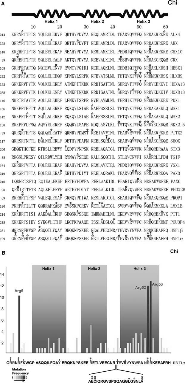
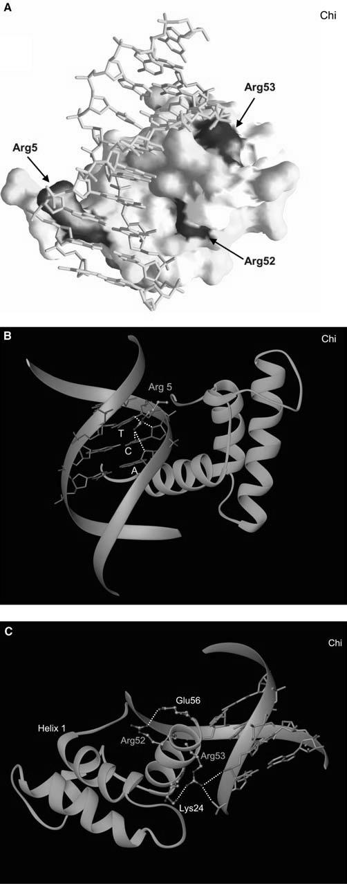
The homeodomain is a highly conserved DNA-binding motif that is found in numerous transcription factors throughout a large variety of species from yeast to humans. These gene-specific transcription factors play critical roles in development and adult homeostasis, and therefore, any germline mutations associated with these proteins can lead to a number of congenital abnormalities. Although much has been revealed concerning the molecular architecture and the mechanism of homeodomain-DNA interactions, the study of disease-causing mutations can further provide us with instructive information as to the role of particular residues in a conserved mode of action. In this paper, I have compiled the homeodomain missense mutations found in various human diseases and re-examined the functional role of the mutational "hot spot" residues in light of the structures obtained from crystallography. These findings should be useful in understanding the essential components of the homeodomain and in attempts to design agonist or antagonists to modulate their activity and to reverse the effects caused by the mutations.
Figures
References
-
- Ades SE, Sauer RT. Differential DNA-binding specificity of the engrailed homeodomain: the role of residue 50. Biochemistry. 1994;33:9187–9194. - PubMed
-
- Aishima J, Wolberger C. Insights into nonspecific binding of homeodomains from a structure of MATalpha2 bound to DNA. Proteins. 2003;51:544–551. - PubMed
-
- Antonarakis SE, Krawczak M, Cooper DN. Disease-causing mutations in the human genome. Eur J Pediatr. 2000;159(Suppl 3):S173–S178. - PubMed
-
- Billeter M. Homeodomain-type DNA recognition. Prog Biophys Mol Biol. 1996;66:211–225. - PubMed
Publication types
MeSH terms
Substances
Grants and funding
LinkOut - more resources
Full Text Sources
Medical
