. 2005 Mar;41(3):535-44.
doi: 10.1002/hep.20600.

Transdifferentiation of rat hepatocytes into biliary cells after bile duct ligation and toxic biliary injury

Affiliations

Transdifferentiation of rat hepatocytes into biliary cells after bile duct ligation and toxic biliary injury

George K Michalopoulos et al. Hepatology. 2005 Mar.

Abstract

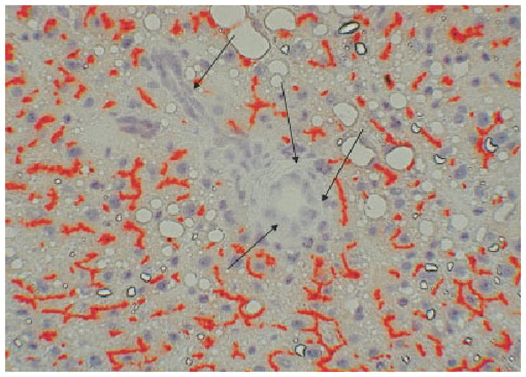
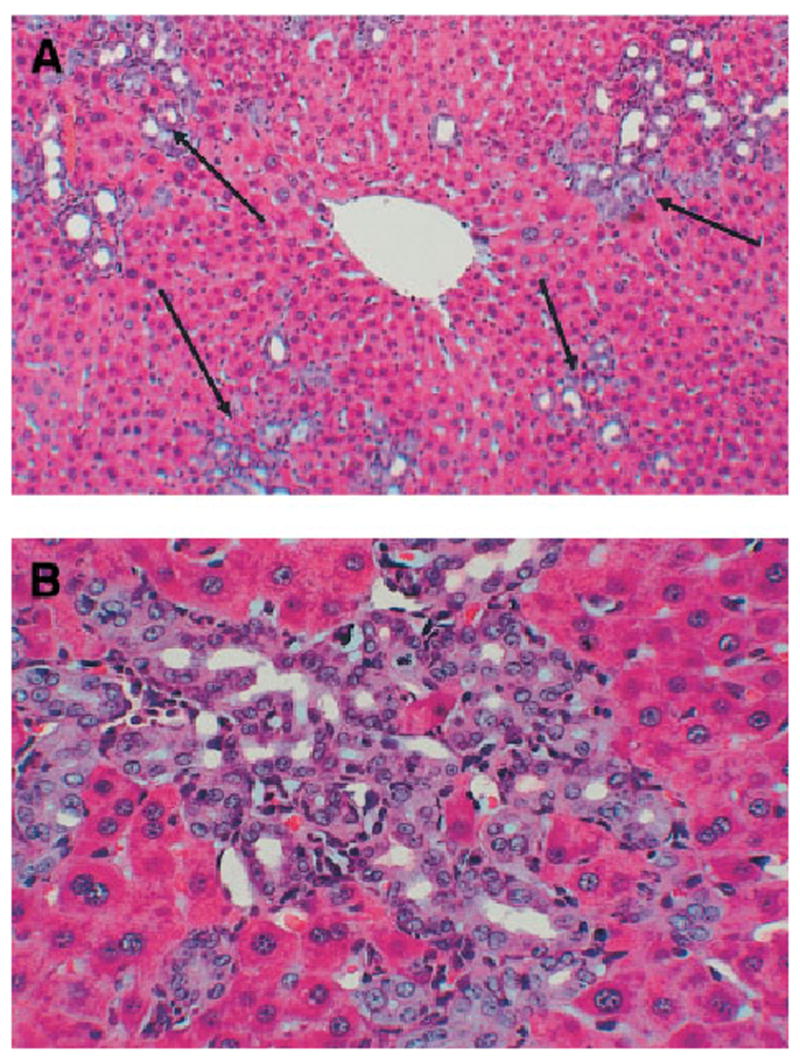
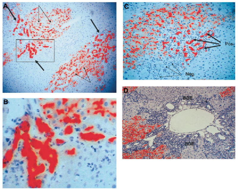
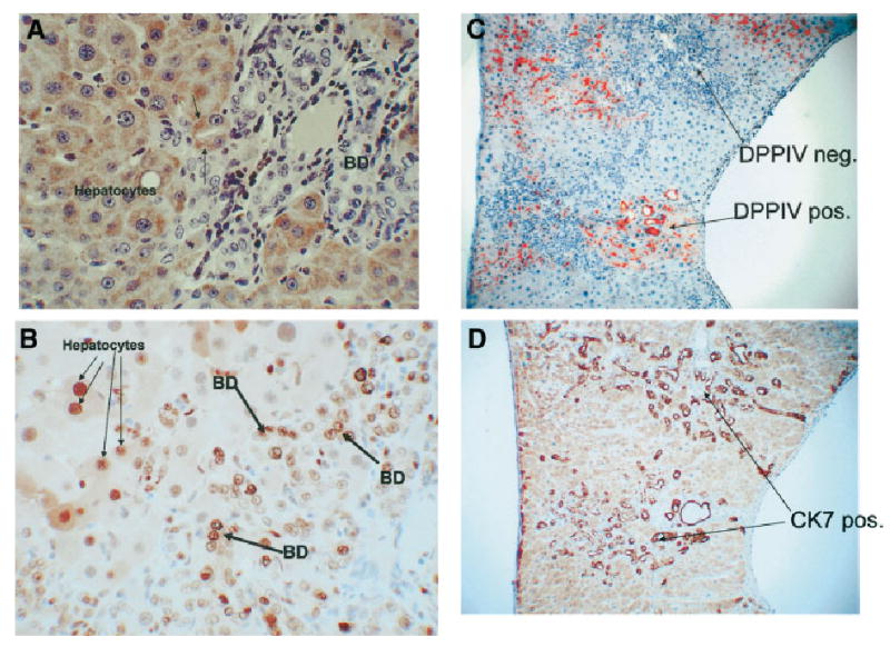
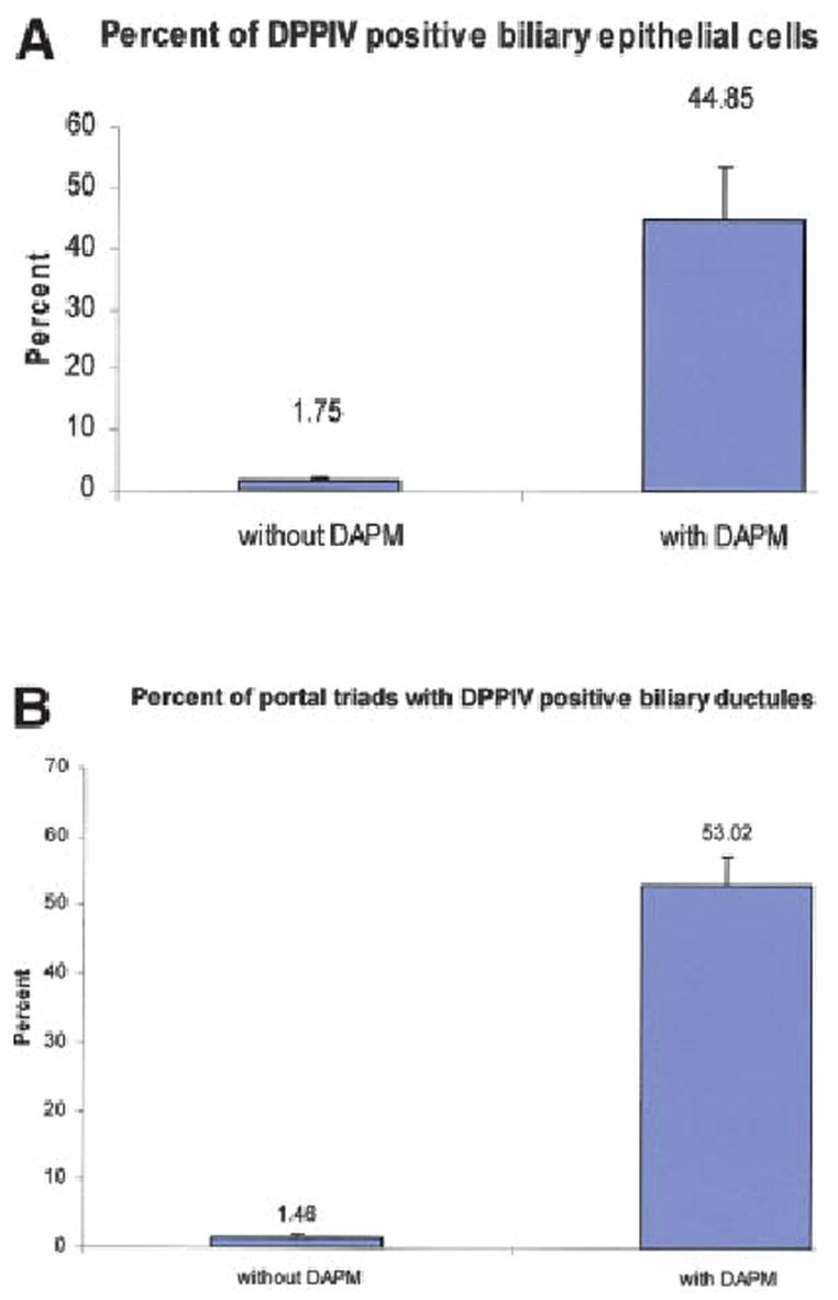
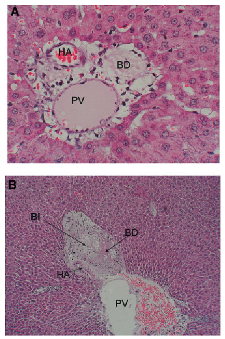
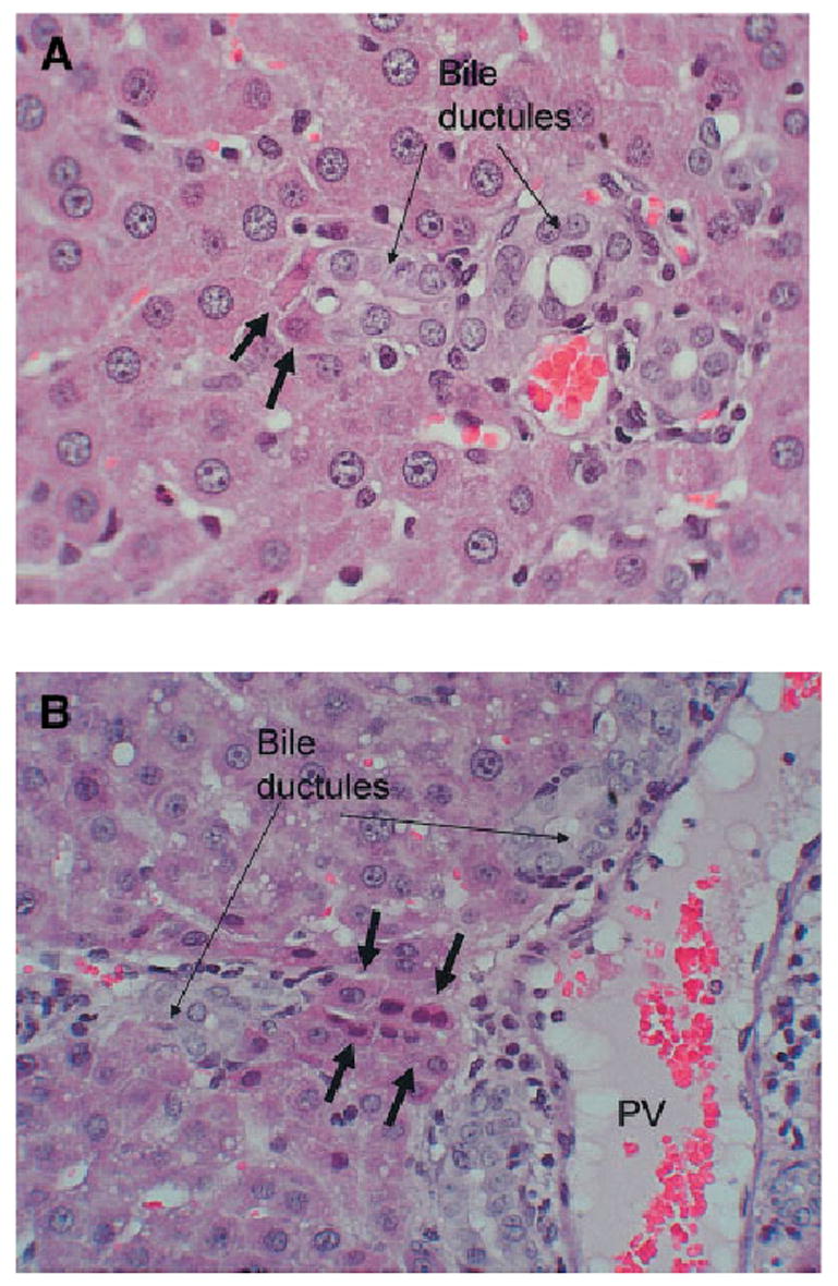
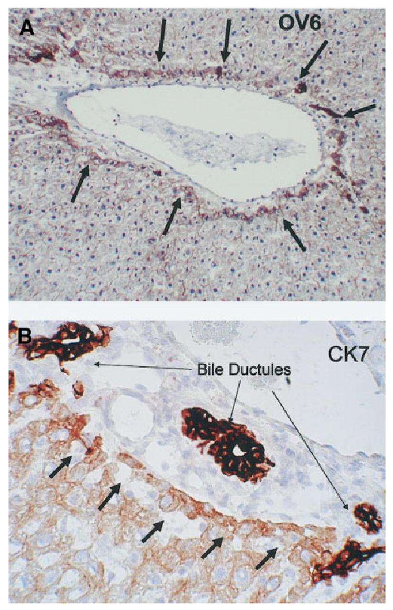
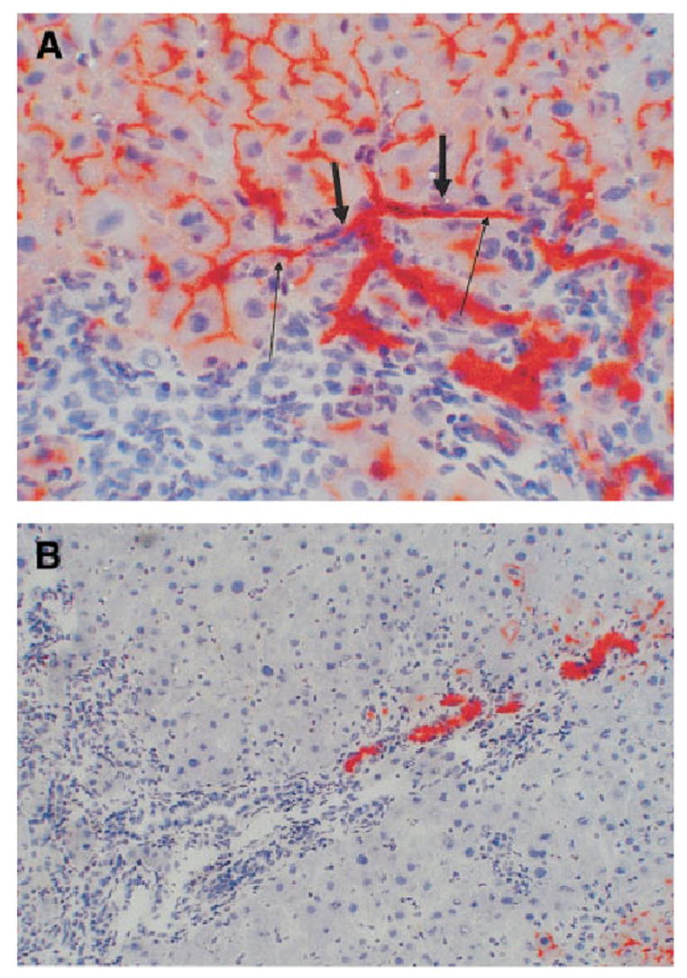
Rats with chimeric livers were generated by using the protocol of injecting hepatocytes from dipeptidyl peptidase IV (DPPIV)-positive donors into retrorsine-treated DPPIV-negative recipients subjected to partial hepatectomy. Rats with established chimeric livers were subjected to bile duct ligation, with or without pretreatment with the biliary toxin methylene diamiline (DAPM). Ductules bearing the donor hepatocyte marker DPPIV were seen at 30 days after bile duct ligation. The frequency of the ductules derived from the donor hepatocytes was dramatically enhanced (36-fold) by the pretreatment with DAPM. In conclusion, our results show that hepatocytes can function as facultative stem cells and rescue the biliary epithelium during repair from injury when its proliferative capacity is being compromised.


Conflict of interest statement

Conflict of interest: Nothing to report.

Figures

Fig. 1
Fig. 1
DPPIV histochemistry of chimeric liver before bile duct ligation. Bile canaliculi of donor hepatocytes stain red (positive) for DPPIV. Bile ductules in portal triads of the recipient liver (arrows) are uniformly negative for DPPIV (original magnification ×200).
Fig. 2
Fig. 2
Hematoxylineosin stain of livers at day 30 after bile duct ligation. (A) Portal triads are expanded with numerous bile ductules (arrows). (original magnification ×200). (B) High-power view of a cluster of bile ductules (original magnification ×400). There was no detectable difference in histology between livers exposed to DAPM versus controls.
Fig. 3
Fig. 3
(A) DPPIV histochemistry shows multiple clusters of DPPIV-positive (red) ductules (thick arrows). The canaliculi of the donor hepatocytes also stain positive for DPPIV (thin arrows) (original magnification ×200). The area surrounded by a square is magnified in panel B (original magnification ×400). (C) A portal triad with a portion of the ductules being DPPIV-positive (red) (thick arrows), whereas another portion has ductules that are DPPIV-negative (thin arrows). (original magnification ×300). (D) A portal triad in which all ductules are negative for DPPIV (original magnification ×200). BDE, bile duct epithelium.
Fig. 4
Fig. 4
A DAPM-BDL protocol at day 30. Immunohistochemical stain for the hepatocyte marker HEPPAR. (A) All hepatocytes stain positive (brown), whereas biliary ductules are negative. (original magnification ×400). Two hepatocytes, however, are arranged in a ductular configuration (arrows). (B) Day 30 of the DAPM-BDL protocol. Immunohistochemical stain for PCNA, indicating presence of cells in the cell cycle. Numerous nuclei of both hepatocytes (thin arrows) and biliary epithelial cells (bile duct, thick arrows) are positive for PCNA. (original magnification ×400). (C–D) Two groups of bile ductules are shown in panel C, of which one is positive and the other negative for DPPIV. Panel D demonstrates that, regardless of the DPPIV expression, both groups of ductules are indistinguishable in terms of CK7. (original magnification ×200). BD, bile duct.
Fig. 5
Fig. 5
Quantification of (A) the percentage of DPPIV-positive bile duct epithelial cells and (B) the percentage of portal triads bearing DPPIV-positive ductules. Data were derived from 3 rats not treated with DAPM and 4 rats treated with DAPM. Three sections were taken randomly from each rat liver. A total of 50 portal triads per rat liver were randomly assessed to derive the data indicated. Each bar represents the mean (± SE) derived by statistical analysis of the data derived from the animals in each group.
Fig. 6
Fig. 6
Histological changes after administration of DAPM. (A) Section of a portal triad at day 2 after administration of DAPM. The bile ductule is devoid of biliary epithelium. (original magnification ×400). (B) An area of hepatocyte necrosis (BI) in immediate proximity to a ruptured bile ductule, at day 4 after DAPM. (original magnification ×100). PV, portal vein branch; HA, hepatic artery branch; BD, interlobular bile ductile; BI, bile infarct.
Fig. 7
Fig. 7
Hematoxylineosin stain of liver sections at day 12 of the DAPM-BDL protocol (original magnification ×200). (A) Two hepatocytes (short arrows) are intercalated into the structure of a bile ductule (long arrows). (B) Hepatocytes are arranged in a ductular configuration (short arrows). PV, portal vein.
Fig. 8
Fig. 8
(A) Immunohistochemistry for OV6. This antibody stains positive in oval cells and biliary epithelium. Hepatocytes immediately adjacent to the portal triad (arrows) become positive for OV56 at day 3 of the DAPM-BDL protocol (original magnification ×100). (B) Day 9 of the DAPM-BDL protocol. Hepatocytes immediately adjacent to the portal triad (short arrows) become moderately positive for CK7. The bile ductules of the same triad (long arrows) are intensely positive for CK7 (original magnification ×200).
Fig. 9
Fig. 9
DAPM-BDL protocol at day 30. (A) Connections between the lumen of the bile ductules (short arrows) and bile canaliculi of hepatocytes (long arrows). (original magnification ×400). (B) DPPIV-positive bile ductules (red) are arranged linearly within a complex of DPPIV-negative ductules (blue-gray) draining into a portal triad (original magnification ×100).

Similar articles

Cited by

References

    1. Alison M, Golding M, Lalani EN, Nagy P, Thorgeirsson S, Sarraf C. Wholesale hepatocytic differentiation in the rat from ductular oval cells, the progeny of biliary stem cells. J Hepatol. 1997;26:343–352. - PubMed
    1. Evarts RP, Nagy P, Marsden E, Thorgeirsson SS. A precursor-product relationship exists between oval cells and hepatocytes in rat liver. Carcinogenesis. 1987;8:1737–1740. - PubMed
    1. Evarts RP, Nagy P, Nakatsukasa H, Marsden E, Thorgeirsson SS. In vivo differentiation of rat liver oval cells into hepatocytes. Cancer Res. 1989;49:1541–1547. - PubMed
    1. Evarts RP, Hu Z, Omori N, Omori M, Marsden ER, Thorgeirsson SS. Precursor-product relationship between oval cells and hepatocytes: comparison between tritiated thymidine and bromodeoxyuridine as tracers. Carcinogenesis. 1996;17:2143–2151. - PubMed
    1. Golding M, Sarraf CE, Lalani EN, Anilkumar TV, Edwards RJ, Nagy P, et al. Oval cell differentiation into hepatocytes in the acetylaminofluorene-treated regenerating rat liver. Hepatology. 1995;22:1243–1253. - PubMed

Publication types