. 2005 Jun 15;105(12):4853-60.
doi: 10.1182/blood-2004-12-4948. Epub 2005 Feb 22.

The hydration state of human red blood cells and their susceptibility to invasion by Plasmodium falciparum

Affiliations

The hydration state of human red blood cells and their susceptibility to invasion by Plasmodium falciparum

Teresa Tiffert et al. Blood. .

Abstract

In most inherited red blood cell (RBC) disorders with high gene frequencies in malaria-endemic regions, the distribution of RBC hydration states is much wider than normal. The relationship between the hydration state of circulating RBCs and protection against severe falciparum malaria remains unexplored. The present investigation was prompted by a casual observation suggesting that falciparum merozoites were unable to invade isotonically dehydrated normal RBCs. We designed an experimental model to induce uniform and stable isotonic volume changes in RBC populations from healthy donors by increasing or decreasing their KCl contents through a reversible K(+) permeabilization pulse. Swollen and mildly dehydrated RBCs were able to sustain Plasmodium falciparum cultures with similar efficiency to untreated RBCs. However, parasite invasion and growth were progressively reduced in dehydrated RBCs. In a parallel study, P falciparum invasion was investigated in density-fractionated RBCs from healthy subjects and from individuals with inherited RBC abnormalities affecting primarily hemoglobin (Hb) or the RBC membrane (thalassemias, hereditary ovalocytosis, xerocytosis, Hb CC, and Hb CS). Invasion was invariably reduced in the dense cell fractions in all conditions. These results suggest that the presence of dense RBCs is a protective factor, additional to any other protection mechanism prevailing in each of the different pathologies.


Figures

Figure 1.
Figure 1.
Linear regression fit of the correlation between cell density and MCHC in normal human RBCs. The points represent data from Table 1, column 2 plotted against column 3. The regression coefficient was r2 = 0.996. The following equation reports the parameters of the fit and can be applied to estimate intermediate and extrapolated data within and outside the measured range: density = 0.00304*MCHC + 0.994.
Figure 2.
Figure 2.
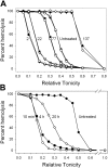
Osmotic fragility curves of K+-equilibrated RBCs. (A) The numbers next to the curves indicate the initial [K+], in mM, in the extracellular media used for K+ equilibration, as described in “Materials and methods” for study I. Open and filled paired symbols report duplicate measurements for each condition. Left shifts relative to the untreated controls indicate RBC dehydration; right shifts indicate RBC swelling. The curves represent integrals of near Gaussian distributions.29,40 Therefore, the approximate parallelism in the observed shifts indicates that the population variation in RBC volumes did not change much in the different hydration states relative to that in the untreated controls, despite the large changes in mean cell volume. (B). Time-dependent shifts in the osmotic fragility curves of markedly dehydrated RBCs (K+ equilibrated in 3 mM [K+]). The curve at 10 minutes approximates the initial osmotic fragility pattern of the dehydrated RBCs at the start of culture. With time (4 hours and 20 hours), there is a right shift in the osmotic fragility curves, reflecting spontaneous rehydration. The progressive decrease in slope indicates increasing population variation of RBC volumes.
Figure 3.
Figure 3.
Morphologic appearance of fresh, unfixed samples of initially profoundly dehydrated RBCs at different times of incubation in culture media. RBCs in culture medium were observed under phase contrast at × 630 (Zeiss photomicroscope III RS; Zeiss Planapo objective lens, ×63 magnification, 1.4 numerical aperture). Photographs were taken with a JVC color video camera, TK-C1381. Note the gradual appearance of smooth, flattened discs, some with typical biconcave shape, indicating spontaneous rehydration of RBCs. This explains the developing heterogeneity apparent in the osmotic fragility curves of Figure 2B.
Figure 4.
Figure 4.
[3H]hypoxanthine incorporation in P falciparum cultures sustained with RBCs in different hydration states. Following parasite invasion in cultures sustained with RBCs of different volumes, parasites were ring-stage synchronized and incubated with [3H]hypoxanthine for 24 hours. After this period, parasite growth was assessed by measuring [3H]hypoxanthine incorporation, as described in “Materials and methods.” The columns and error bars report the means and SDs of quadruplicate measurements expressed as a percent of the incorporation in untreated controls (right column).
Figure 5.
Figure 5.
[3H]hypoxanthine incorporation after 4 hours and 24 hours of incubation of P falciparum cultures sustained with RBCs in different hydration states. The experimental protocol was similar to that indicated in Figure 4 except that [3H]hypoxanthine incorporation was measured at 4 hours (A), representing invasion efficiency, and 24 hours (B), reflecting invasion and parasite growth. Note the orders of magnitude difference in the ordinate scales between panels A and B. Panel C reports the [3H]hypoxanthine incorporation ratio (24 hours-4 hours)/4 hours, estimated from randomly paired samples. This ratio is assumed to reflect mainly parasite growth rate because the extent of [3H]hypoxanthine incorporation attributed to invasion at 4 hours is subtracted in the calculations. The right columns in each panel report [3H]hypoxanthine incorporation in untreated controls. Columns and error bars represent means and SDs of triplicate measurements.
Figure 6.
Figure 6.
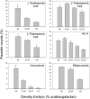
P falciparum invasion efficient in normal RBCs of different densities. Normal RBCs from 4 different donors were harvested from the indicated arabinogalactan concentration ranges and used to sustain P falciparum cultures as described in “Materials and methods.” Parasite counts were assessed on Giemsa-stained blood smears after 38 hours of incubation of the culture. At least 1000 RBCs were counted per slide on 3 different slides. “All” indicates parasite counts in the original, unfractionated RBC samples. The columns and error bars report the means and SDs of the triplicate counts. Labels 1-4 in quadrants indicate a different donor.
Figure 7.
Figure 7.
P falciparum invasion efficiency in density-separated RBCs with various congenital abnormalities. RBCs from 6 different donors with the indicated pathologies were harvested from the arabinogalactan concentration ranges shown on the abscissa and used to sustain P falciparum cultures in conditions similar to those indicated in Figure 6. “All” indicates parasite counts in the original, unfractionated RBC samples. Parasite counts were assessed after 38 hours of incubation on Giemsa-stained blood smears. At least 1000 RBCs were counted per slide on 3 different slides. The columns and error bars report the means and SDs of these triplicate counts.

References

    1. Dvorak JA, Miller LH, Whitehouse WC, Shiroishi T. Invasion of erythrocytes by malaria merozoites. Science. 1975;187: 748-750. - PubMed
    1. Bannister LH, Dluzewski AR. The ultrastructure of red cell invasion in malaria infections: a review. Blood Cells. 1990;16: 257-292. - PubMed
    1. Wilson RJM. Biochemistry of red cell invasion. Blood Cells. 1990;16: 237-252. - PubMed
    1. Berzins K. Merozoite antigens involved in invasion. Chem Immunol. 2002;80: 125-143. - PubMed
    1. Gaur D, Mayer DC, Miller LH. Parasite ligand-host receptor interactions during invasion of erythrocytes by Plasmodium merozoites. Int J Parasitol. 2004;34: 1413-1429. - PubMed

Publication types

MeSH terms