

Derivation of 2 categories of plasmacytoid dendritic cells in murine bone marrow
- PMID: 15728131
- PMCID: PMC1850236
- DOI: 10.1182/blood-2004-07-2529
Derivation of 2 categories of plasmacytoid dendritic cells in murine bone marrow
Abstract
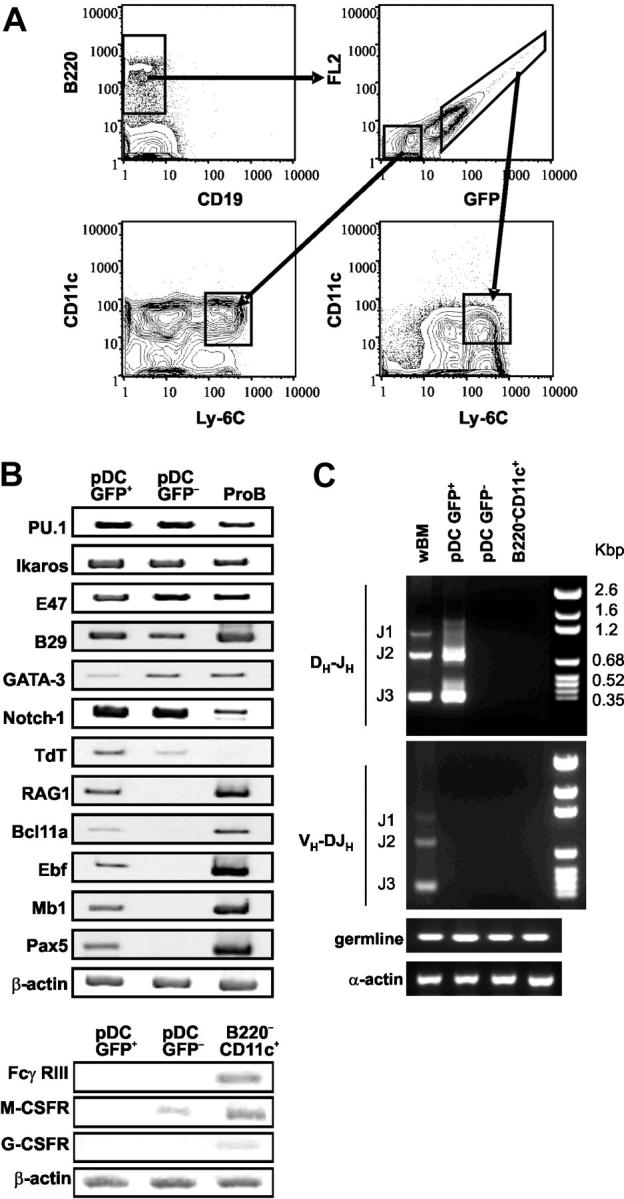
Plasmacytoid dendritic cells (pDCs) competent to make type I interferon were rigorously defined as a Ly-6C(+) and CD11c(Lo) subset of the B220(+)CD19(-) CD43(+)CD24(Lo) bone marrow (BM) Fraction A. Otherwise similar Ly6C(-) cells expressed the natural killer (NK) markers DX5 and NK1.1. pDCs represented a stable, discrete, and long-lived population. Stem cells and early lymphoid progenitors (ELPs), but not prolymphocytes, were effective precursors of pDCs, and their differentiation was blocked by ligation of Notch receptors. Furthermore, pDCs were present in the BM of RAG1(-/-), CD127/IL-7Ra(-/-), and Pax5(-/-) mice. pDCs in RAG1/GFP knock-in mice could be subdivided, and immunoglobulin D(H)-J(H) rearrangements, as well as transcripts for the B-lineage-related genes Pax5, mb1/CD79a, ebf, and Bcl11a, were identified only in the green fluorescent protein-positive (GFP(+)) pDC1 subset. All pDCs expressed terminal deoxynucleotidyl transferase (TdT), the ETS transcription factor Spi-B, the nuclear factor-kappaB transcription factor RelB, toll-like receptor 9 (TLR9), and interferon consensus sequence binding protein (ICSBP)/interferon regulatory factor 8 (IRF-8) transcripts; lacked CD16 and granulocyte colony-stimulating factor receptor (G-CSFR); and were uniformly interleukin-7 receptor alpha (IL-7Ralpha(-)) AA4.1(Lo), CD27(-), Flk-2(Lo), c-Kit(-), DX-5(-), and CD11b(-), while CD4 and CD8alpha were variable. GFP(+) pDC1 subset was less potent than GFP(-) pDC2s in T allostimulation and production of tumor necrosis factor alpha (TNFalpha), interferon alpha (IFNalpha), and interleukin-6 (IL-6), while only pDC2s made IFNgamma and IL-12 p70. Thus, 2 functionally specialized subsets of pDCs arise in bone marrow from progenitors that diverge from B, T, and NK lineages at an early stage.
Figures
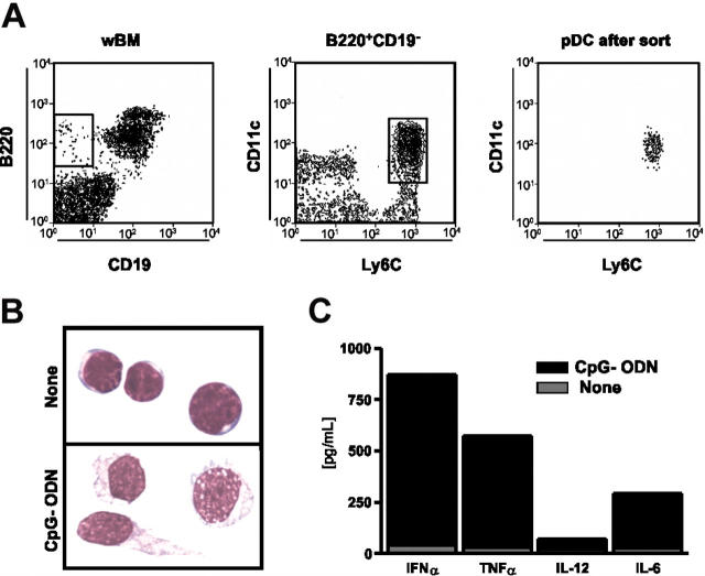
 ). These results are representative of those obtained in 4 independent experiments.
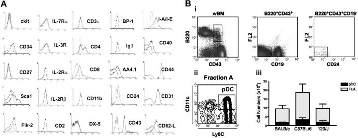 ) recovered from 4 harvested bones from 3 strains of mice are shown in the bar graph. Results are presented as the mean ± SD of 5 different experiments.
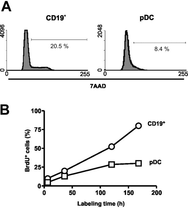


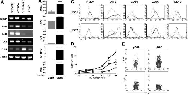 , pDC1; ▪, pDC2) were stimulated for 12 hours with CpG-ODNs. Supernatants were analyzed for IFNα, TNFα, IL-6, IL-12p70, and IFNγ levels by ELISA. Results are presented as the mean ± SD of 3 different experiments. (C) Expression of costimulatory molecules was assessed on pDC1s and pDC2s after 18-hour stimulation with CpG-ODNs. Dotted lines indicate unstimulated cells. (D) Preactivated RAG1+ pDC1s (□), RAG1- pDC2s (○), and B220-CD11c+ DCs (▵) were cultured for 6 days with 1 × 10⁵ allogeneic T cells. Proliferation of TCRβ+ cells was measured by BrdU incorporation as described in “Material and methods.” Of 3 experiments, 1 representative is shown. Results are presented as the mean ± SD of 3 different culture wells; one representative experiment of 3 is shown. (E) Intracellular cytokine accumulation was determined in T cells 6 days after priming with preactivated pDC1s or pDC2s, and 5-hour restimulation with PMA and ionomycin.
References
-
- Asselin-Paturel C, Boonstra A, Dalod M, et al. Mouse type I IFN-producing cells are immature APCs with plasmacytoid morphology. Nat Immunol. 2001;2: 1144-1150. - PubMed
-
- Blanco P, Palucka AK, Gill M, Pascual V, Banchereau J. Induction of dendritic cell differentiation by IFN-α in systemic lupus erythematosus. Science. 2001;294: 1540-1543. - PubMed
-
- Jego G, Palucka AK, Blanck JP, et al. Plasmacytoid dendritic cells induce plasma cell differentiation through type I interferon and interleukin 6. Immunity. 2003;19: 225-234. - PubMed
Publication types
MeSH terms
Substances
Grants and funding
LinkOut - more resources
Full Text Sources
Other Literature Sources
Medical
Research Materials
Miscellaneous
