. 2005 Feb 28;168(5):813-24.
doi: 10.1083/jcb.200406083. Epub 2005 Feb 22.

The LIM protein Ajuba influences p130Cas localization and Rac1 activity during cell migration

Affiliations

The LIM protein Ajuba influences p130Cas localization and Rac1 activity during cell migration

Stephen J Pratt et al. J Cell Biol. .

Abstract

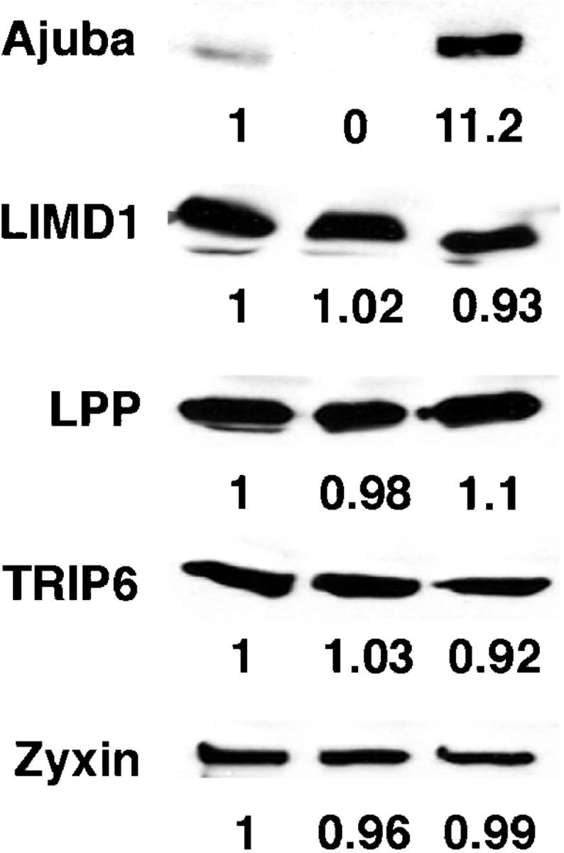
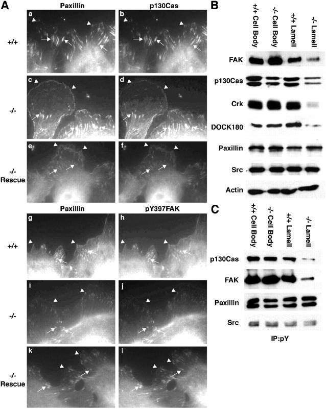
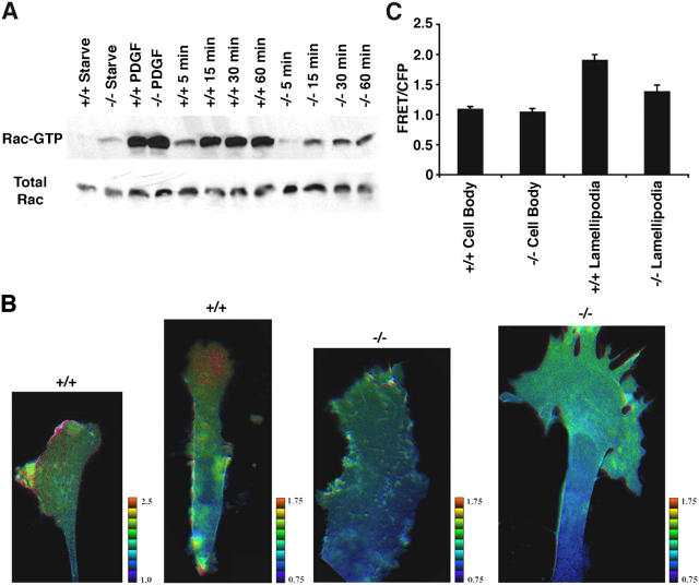
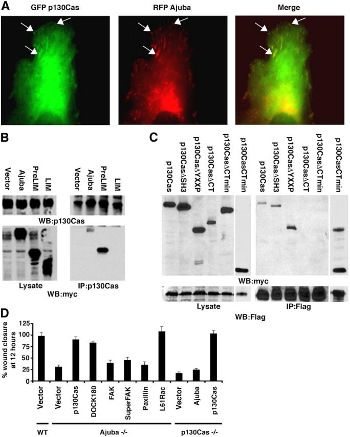
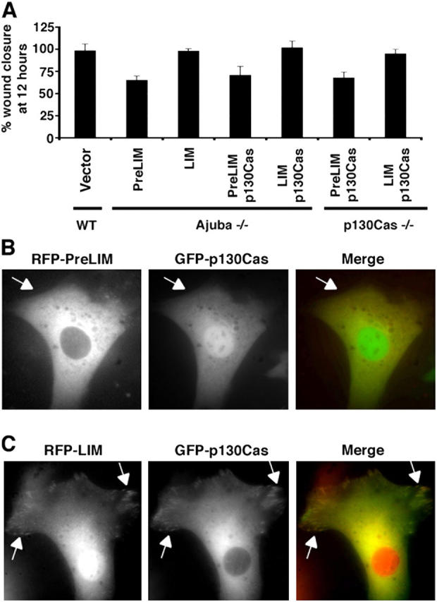
Cell migration requires extension of lamellipodia that are stabilized by formation of adhesive complexes at the leading edge. Both processes are regulated by signaling proteins recruited to nascent adhesive sites that lead to activation of Rho GTPases. The Ajuba/Zyxin family of LIM proteins are components of cellular adhesive complexes. We show that cells from Ajuba null mice are inhibited in their migration, without associated abnormality in adhesion to extracellular matrix proteins, cell spreading, or integrin activation. Lamellipodia production, or function, is defective and there is a selective reduction in the level and tyrosine phosphorylation of FAK, p130Cas, Crk, and Dock180 at nascent focal complexes. In response to migratory cues Rac activation is blunted in Ajuba null cells, as detected biochemically and by FRET analysis. Ajuba associates with the focal adhesion-targeting domain of p130Cas, and rescue experiments suggest that Ajuba acts upstream of p130Cas to localize p130Cas to nascent adhesive sites in migrating cells thereby leading to the activation of Rac.


Figures

Figure 1.
Figure 1.
Quantitative Western blot analyses of Zyxin/Ajuba family members. WT (+/+, lane 1), Ajuba null (−/−, lane 2), and Ajuba rescued Ajuba null MEFs (−/− Rescue, lane 3). Equal amounts of protein were loaded in each lane. Densitometry was performed and the level of each protein is displayed below each lane. The wt sample is arbitrarily set at 1.
Figure 2.
Figure 2.
Migration of primary MEFs from Ajuba null mice is inhibited. (A) Phase-contrast images of scratch-wound assays of WT and Ajuba null (−/−) MEFs over a 10-h time course. (B) Stick figure of Ajuba and the NH2-terminal PreLIM region and COOH-terminal LIM region. Percent wound closure at 12 h of wt MEFs (+/+, column 1) or Ajuba null MEFs (−/−, columns 2–6) transfected with: RFP alone (+RFP), full-length Ajuba (+RFP-Ajuba), the PreLIM region of Ajuba (+RFP-PreLIM), the LIM region of Ajuba (+RFP-LIM), or both PreLIM and LIM regions of Ajuba expressed by separate plasmids (+GFP-PreLIM and RFP-LIM). WT cells are arbitrarily set at 100%. Multiple wounds were examined and data presented as the mean value plus the SD about the mean.
Figure 3.
Figure 3.
Ajuba null MEFs adhere to ECM proteins, spread, and activated integrin receptors normally. (A) Cell adhesion. WT (♦) and Ajuba null (□) MEFs were allowed to adhere for 1 h to plates coated with increasing concentration of the indicated ECM protein. (B) Cell spreading. WT (♦) and Ajuba null (□) MEFs were allowed to adhere and spread on fibronectin-coated coverslips over 2 h. Videos of multiple cells were obtained and data extrapolated at the indicated time points. Results are the mean values plus the SD about the mean. (C) Serum-starved wt (+/+) and Ajuba null (−/−) cells were allowed to adhere to fibronectin-coated plates for 1 h. (D) FAK and p130Cas were immunoprecipitated from cells in suspension (Sus) or bound to fibronectin (Fib) and bound products Western blotted with anti-pY397FAK or anti-phosphotyrosine antibodies, respectively. Actin Western blots are loading controls.
Figure 4.
Figure 4.
Lamellipodia production by migrating Ajuba null MEFs was abnormal. Phalloidin staining of multiple wt (A) and Ajuba null (−/−) (B) MEFs at the wound edge. The direction of migration is toward the top of the page. Arrows identify ruffles/lamellipodia. Both images were obtained using a 20× lens NA 1.4. (C) Quantitative lamellipodia assay as described in Materials and methods. To control for random lamellipodia production control filters were coated with BSA at each time point. Pseudopodia/lamellipodia production versus time of serum stimulation is presented as protein produced (OD562). Error bars represent the SD from the mean of multiple experiments.
Figure 5.
Figure 5.
FAK and p130Cas, but not paxillin, protein levels and tyrosine phosphorylation were decreased at focal complexes in migrating Ajuba null lamellipodia. (A) Indirect immunofluorescence of migrating wt (+/+; a, b, g, h), Ajuba null (−/−; c, d, i, j), and Ajuba rescued Ajuba null MEFs (−/− Rescue; e, f, k, l) at the wound edge. Cells were costained for p130Cas (b, d, f) or pY397-FAK (h, j, l), and paxillin (a, c, e, g, i, k). Secondary antibodies conjugated with FITC (p130Cas and pY397FAK) or Alexa Fluor 568. Arrowheads identify leading edge focal complexes whereas arrows identify focal adhesions present at the base of the lamellipodia. Cells were imaged using a 60× oil immersion lens (NA 1.4). (B) Western blot analysis for the indicated proteins was performed on lysates of cell bodies and lamellipodia from wt (+/+) and Ajuba null (−/−) MEFs. Samples were normalized so that an equal amount of protein was loaded in each lane. (C) Phosphotyrosine-containing proteins were immunoprecipitated from lysates of wt (+/+) and Ajuba null (−/−) cell bodies and lamellipodia and bound products Western blotted for the indicated proteins. Each sample was normalized to equal amounts of protein before immunoprecipitation.
Figure 6.
Figure 6.
Migration induced Rac activation is reduced in Ajuba null MEFs. (A) WT (+/+, lanes 1, 3, 5–8) or Ajuba null (−/−, lanes 2, 4, 9–12) MEFs were starved overnight (lanes 1 and 2), stimulated with PDGF (lanes 3 and 4) or scratch wounded for the indicated times (lanes 5–12). Rac Western blots for GTP-bound Rac pull downs (top) and total Rac (bottom). (B) Rac-FRET analysis of migrating wt (+/+) and Ajuba null (−/−) MEFs. Cells are migrating toward the top of the page. Red and blue indicate high and low FRET activity, respectively. Scales for each image are shown to the right. (C) Quantification of FRET activity. Average FRET/CFP pixel intensity of the leading edge versus cell body in wt (+/+) versus Ajuba null (−/−) MEFs are shown. 30 or more cells were analyzed for each sample.
Figure 7.
Figure 7.
Ajuba colocalized with p130Cas at focal complexes and focal adhesions in lamellipodia, and associated with p130Cas in cells. (A) WT MEFs were cotransfected with GFP-p130Cas (green) and RFP-Ajuba (red) and induced to migrate (toward top of page) by scratch wounding. Arrows indicate localization of p130Cas and Ajuba (colocalization, yellow in merged image). (B) COS-7 cells were transfected with myc-tagged Ajuba plasmids, endogenous p130Cas was immunoprecipitated (right) and bound products Western blotted for the presence of Ajuba (anti-myc; bottom) and p130Cas (top). Left panels are loading controls of cell lysates. (C) COS-7 cells were cotransfected with Flag-tagged Ajuba and various myc-tagged p130Cas mutants. Ajuba was immunoprecipitated (anti-Flag; right) and bound products Western blotted for the presence of p130Cas (anti-myc; top) and Ajuba (anti-Flag; bottom). Loading controls of cell lysates (left). (D) Migration rescue experiments. WT, Ajuba −/−, and p130Cas −/− MEFs were transfected with control empty GFP vector, RFP-Ajuba, GFP-p130Cas, GFP-DOCK180, GFP-paxillin, SuperFAK-IRES.GFP, or myc-Rac1(L61) as indicated. Confluent cell layers were scratch wounded, and videos of wound repair obtained, as described in Fig. 2 B. Shown is the percent wound closure at 12 h. Multiple wounds were analyzed and the data presented as the mean and the SD about the mean.
Figure 8.
Figure 8.
PreLIM Ajuba blocks p130Cas localization to adhesive sites and p130 rescue of migration. (A) Migration rescue experiments. WT, Ajuba −/−, and p130Cas −/− MEFs were transfected with control empty GFP vector, RFP-PreLIM, RFP-LIM, both RFP-PreLIM and GFP-p130Cas, or both RFP-LIM and GFP-p130Cas as indicated. Confluent cell layers were scratch wounded, and videos of wound repair were obtained, as described in Fig. 2 B. Shown is the percent wound closure at 12 h. Multiple wounds were analyzed and the data presented as the mean and the SD about the mean. (B and C) Cells were cotransfected with RFP-PreLIM and GFP-p130Cas (B), or RFP-LIM and GFP-p130Cas (C), and immunofluorescence performed. Arrows identify focal adhesions.

Similar articles

Cited by

References

    1. Bear, J.E., T.M. Svitkina, M. Krause, D.A. Schafer, J.J. Loureiro, G.A. Strasser, I.V. Maly, O.Y. Chaga, J.A. Cooper, G.G. Borisy, and F.B. Gertler. 2002. Antagonism between Ena/VASP proteins and actin filament capping regulates fibroblast motility. Cell. 109:509–521. - PubMed
    1. Beningo, K.A., M. Dembo, I. Kaverina, J.V. Small, and Y.L. Wang. 2001. Nascent focal adhesions are responsible for the generation of strong propulsive forces in migrating fibroblasts. J. Cell Biol. 153:881–888. - PMC - PubMed
    1. Bouton, A.H., R.B. Riggins, and P.J. Bruce-Staskal. 2001. Functions of the adapter protein Cas: signal convergence and the determination of cellular responses. Oncogene. 20:6448–6458. - PubMed
    1. Brown, M.C., K.A. West, and C.E. Turner. 2002. Paxillin-dependent paxillin kinase linker and p21-activated kinase localization to focal adhesions involves a multistep activation pathway. Mol. Biol. Cell. 13:1550–1565. - PMC - PubMed
    1. Brugnera, E., L. Haney, C. Grimsley, M. Lu, S.F. Walk, A.C. Tosello-Trampont, I.G. Macara, H. Madhani, G.R. Fink, and K.S. Ravichandran. 2002. Unconventional Rac-GEF activity is mediated through the Dock180-ELMO complex. Nat. Cell Biol. 4:574–582. - PubMed

Publication types

MeSH terms