. 2005 Feb 28;168(5):825-36.
doi: 10.1083/jcb.200408157. Epub 2005 Feb 22.

Molecular dissection of the photoreceptor ribbon synapse: physical interaction of Bassoon and RIBEYE is essential for the assembly of the ribbon complex

Affiliations

Molecular dissection of the photoreceptor ribbon synapse: physical interaction of Bassoon and RIBEYE is essential for the assembly of the ribbon complex

Susanne tom Dieck et al. J Cell Biol. .

Abstract

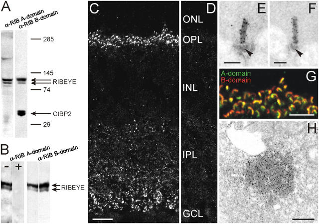
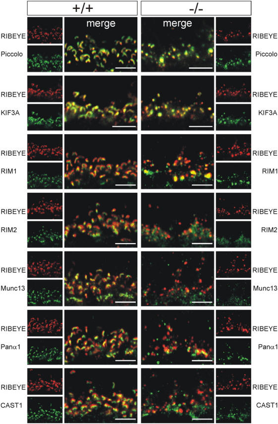
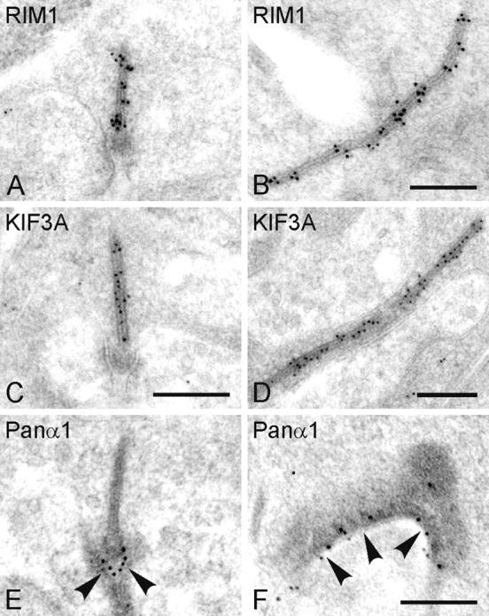
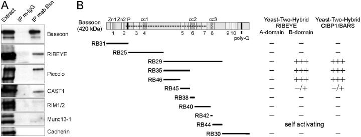
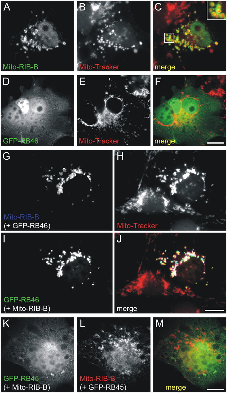
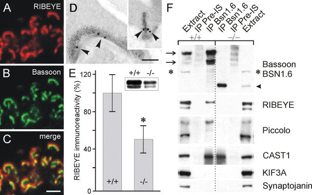
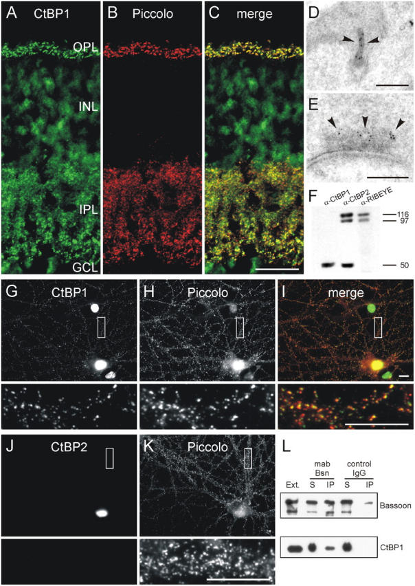
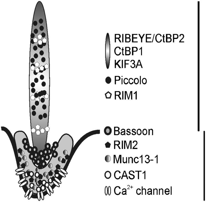
The ribbon complex of retinal photoreceptor synapses represents a specialization of the cytomatrix at the active zone (CAZ) present at conventional synapses. In mice deficient for the CAZ protein Bassoon, ribbons are not anchored to the presynaptic membrane but float freely in the cytoplasm. Exploiting this phenotype, we dissected the molecular structure of the photoreceptor ribbon complex. Identifiable CAZ proteins segregate into two compartments at the ribbon: a ribbon-associated compartment including Piccolo, RIBEYE, CtBP1/BARS, RIM1, and the motor protein KIF3A, and an active zone compartment including RIM2, Munc13-1, a Ca2+ channel alpha1 subunit, and ERC2/CAST1. A direct interaction between the ribbon-specific protein RIBEYE and Bassoon seems to link the two compartments and is responsible for the physical integrity of the photoreceptor ribbon complex. Finally, we found the RIBEYE homologue CtBP1 at ribbon and conventional synapses, suggesting a novel role for the CtBP/BARS family in the molecular assembly and function of central nervous system synapses.


Figures

Figure 1.
Figure 1.
Characterization of the photoreceptor ribbon protein RIBEYE in wild-type and Bassoon mutant mice. (A) Immunoblot of mouse retinal homogenate probed with antibodies against RIBEYE A- and B-domain. Two protein bands (double arrow) of ∼110 kD and 120 kD, representing RIBEYE, are recognized by both antibodies. In addition, the anti-RIBEYE B-domain antibody labels the 50-kD CtBP2 protein band (arrow). Protein bands above and below the RIBEYE doublet are unspecific and appeared only occasionally. (B) Preincubation of the anti-GST-RIBEYE A-domain antiserum with an excess of the MBP-RIB179-448 fusion protein (+) completely prevents the immunodetection of RIBEYE. −, no RIBEYE antigen included. Incubation of the same blot with the anti-RIBEYE B-domain antibody shows the presence of RIBEYE in both lanes. (C) In a vertical cryostat section through mouse retina, the anti-RIBEYE A-domain antiserum stains the photoreceptor ribbons in the outer plexiform layer (OPL) and the bipolar cell ribbons in the inner plexiform layer (IPL). (D) Preadsorption of the anti-RIBEYE antiserum with the antigen results in a complete loss of RIBEYE staining. (E and F) EM and postembedding immunogold labeling shows that photoreceptor ribbons are decorated with gold particles for RIBEYE A- (E) and B-domain (F). Note the absence of gold particles at the base of the ribbons (arrowheads), the region of the arciform density. (G) Confocal laser-scanning micrograph of a region of the OPL double labeled for RIBEYE A- (green) and B-domain (red) demonstrate the colocalization of the two immunoreactivities at the photoreceptor ribbons. (H) An aggregate of free-floating ribbons in a cone photoreceptor terminal of Bassoon mutant retina decorated with RIBEYE immunoreactivity (preembedding labeling). ONL, outer nuclear layer; INL, inner nuclear layer; GCL, ganglion cell layer. Bars: 10 μm (C and D); 0.1 μm (E and F); 5 μm (G); and 0.4 μm (H).
Figure 2.
Figure 2.
Screening the photoreceptor ribbon synaptic complex for ribbon-associated proteins in wild-type (+/+) and Bassoon mutant (−/−) retinae. (Left) Confocal laser-scanning micrographs of photoreceptor ribbons in the +/+ retina double labeled for RIBEYE combined with the following CAZ proteins: Piccolo, KIF3A, RIM1, RIM2, Munc13-1, Ca2+ channel α1 subunit, and ERC2/CAST1. All CAZ proteins colocalize with RIBEYE as seen in the merge of the stainings. (Right) In the −/− retina, Piccolo, KIF3A, and RIM1 colocalize with RIBEYE, but not RIM2, Munc13-1, Ca2+ channel α1 subunit, and ERC2/CAST1. Bars, 5 μm.
Figure 3.
Figure 3.
Postembedding immunogold localization of CAZ proteins at wild-type photoreceptor ribbons. Electron micrographs showing different planes of section through photoreceptor ribbons that are postembedding immunogold labeled for RIM1 (A and B), KIF3A (C and D), and a Ca2+ channel α1 subunit (E and F). The gold particles for RIM1 and KIF3A are associated with the ribbon, the gold particles for the Ca2+ channel α1 subunit are associated with the active zone at the ribbon (arrowheads). Bars, 0.2 μm.
Figure 4.
Figure 4.
Biochemical and yeast two-hybrid analysis of the Bassoon interaction with members of the CtBP family. (A) Immunoblots showing that Bassoon complexes containing RIBEYE, Piccolo, and ERC2/CAST1 can be coimmunoprecipitated from wild-type retina extracts with a monoclonal anti-Bassoon antibody. Under the conditions used, no considerable amounts of RIM1/2, Munc13-1, and cadherin are detectable in immunoprecipitates. (B) Yeast two-hybrid analysis shows strong binding of Bassoon constructs harboring aa 1653–2087 (fragments RB29, RB35, RB46) with the RIBEYE B-domain and the RIBEYE homologue CtBP1. Construct RB45 shows only a weak interaction; construct RB44 is self activating. Specificity of the binding is shown by the lack of binding to the RIBEYE A-domain. −, no reporter gene activation; −/+, weak reporter gene activation; +++, strong reporter gene activation. Zn1, Zn2: double zinc fingers; cc1, cc2, cc3: coiled-coil domains; poly-Q: polyglutamine stretch. Numbers indicate Piccolo–Bassoon homology regions 1–10; dashed line indicates the region of Bassoon missing in the mutant protein.
Figure 5.
Figure 5.
Reconstitution of Bassoon–RIBEYE protein complexes by cotransfection of the Bassoon and RIBEYE binding domains in COS cells. (A–C) Mito-RIBEYE B-domain constructs (A; anti-flag immunosignal) localize to mitochondria (B; MitoTracker Red CMXRos), best seen in the inset in the merge of the two stainings in C. (D–F) The GFP-RB46 (D) construct is distributed diffusely when expressed alone and shows no similarity in pattern with mitochondria (E and F). (G–J) In double-transfection experiments with the Mito-RIB-B-domain, the GFP-RB46 construct (I) adopts a pattern that overlaps with that of the Mito-RIB-B-domain (G) and the MitoTracker (H), as clearly seen when images are merged (J). (K–M) In contrast, the GFP-RB45 construct (K) remains diffusely distributed and shows no enrichment at mitochondria in cells that are cotransfected with the Mito-RIB-B-domain (L), as seen in the merge of the two stainings (M). Bars, 10 μm.
Figure 6.
Figure 6.
Bassoon–RIBEYE interaction at the base of the photoreceptor ribbon. (A–C) Confocal laser-scanning micrographs of photoreceptor ribbon synapses double labeled for RIBEYE (A) and Bassoon (B). In the merge of the two stainings (C), the horseshoe-like appearance of the photoreceptor ribbon colabeled for RIBEYE and Bassoon is clearly visible. Bassoon labeling is surrounded by the RIBEYE labeling. (D) Electron micrographs of photoreceptor ribbons, en face and cross-section view (inset), postembedding immunogold double labeled for Bassoon (large gold particles) and RIBEYE (small gold particles). The gold particles for Bassoon are located closest to the active zone (arrowheads). (E) Immunoblots of 25 μg total protein per lane from wild-type (+/+) and Bassoon mutant (−/−) retina homogenates show a 50% reduction of RIBEYE immunoreactivity in the −/− retina. Data are normalized to the mean value (100%) obtained from wild-type samples. Statistical analysis by the unpaired t test shows a significant difference in the RIBEYE immunoreactivity between the two genotypes (asterisk, P < 0,01). Results are expressed as mean ± SD; n = 4 for both genotypes. (F) Immunoprecipitation experiments from wild-type (+/+) and mutant (−/−) retinal extracts with the rabbit antiserum BSN1.6 directed against the NH2-terminal part of wild-type and mutant Bassoon. The arrows on the left indicate wild-type Bassoon (420 and 320 kD), the arrowhead on the right indicates the 180-kD mutant protein. Note that the unspecific band (asterisk) is not precipitated. RIBEYE is coimmunoprecipitated from wild-type but not from Bassoon mutant retina. The same is true for the higher molecular weight band of Piccolo. In contrast, ERC2/CAST1 is found in both precipitates. Kinesin KIF3A and synaptojanin are not detected in either precipitate. Control experiments were performed with preimmune serum (preIS). IP, immunoprecipitation. Bars: 2 μm (A–C), 0.2 μm (D).
Figure 7.
Figure 7.
CtBP1, a RIBEYE homologue, is expressed at ribbon and conventional synapses. (A–C) Confocal laser-scanning micrographs of a vertical section through mouse retina double labeled for CtBP1 (A) and Piccolo (B), which marks ribbon and conventional synapses in the retina. As seen in the merge of the two stainings (C), CtBP1 and Piccolo immunoreactivities completely colocalize at the synapses in the outer plexiform layer (OPL) and inner plexiform layer (IPL) of the retina. (D and E) Electron micrographs of a photoreceptor (D) and an amacrine cell synapse (E) immunogold labeled for CtBP1. At the ribbon synapse the gold particles for CtBP1 decorate the ribbon (arrowheads), at the amacrine cell synapse they are located some distance from the active zone (arrowheads) at the edge of the electron-dense CAZ material. (F) Western blots of retina homogenate probed with antibodies against CtBP1 and the RIBEYE A- and CtBP2/RIBEYE B-domain. The antibody against CtBP1 recognizes the 50-kD CtBP1 protein and does not cross-react with RIBEYE. (G–I) Micrographs of cultured hippocampal neurons double labeled for CtBP1 (G) and Piccolo (H). In hippocampal neurons, like in retinal neurons, a fraction of CtBP1 immunoreactivity is localized at synapses where it colocalizes with Piccolo as seen in the merge of the two stainings (I). (J and K) CtBP2 (J) is not present at hippocampal synapses labeled with Piccolo (K). (L) Immunoblots showing that CtBP1 can be coimmunoprecipitated from brain synaptosomes with a monoclonal anti-Bassoon antibody. The boxes in G–K mark the regions that are shown at higher magnification. INL, inner nuclear layer; GCL, ganglion cell layer; Ext., brain synaptosomal extract; IP, immunoprecipitate; S, supernatant. Bars: 20 μm (A–C), 0.2 μm (D and E), and 10 μm (G–K).
Figure 8.
Figure 8.
Differential localization of CAZ proteins defines two compartments of the photoreceptor ribbon synaptic complex. The ribbon-associated complex of CAZ proteins includes RIBEYE/CtBP2, CtBP1, KIF3A, Piccolo, and RIM1; the plasma membrane/arciform density-associated complex includes RIM2, Munc13-1, ERC2/CAST1, and a Ca2+ channel α1 subunit. Bassoon localizes at the border between the two compartments.

References

    1. Altrock, W.D., S. tom Dieck, M. Sokolov, A.C. Meyer, A. Sigler, C. Brakebusch, R. Fässler, K. Richter, T.M. Boeckers, H. Potschka, et al. 2003. Functional inactivation of a fraction of excitatory synapses in mice deficient for the active zone protein Bassoon. Neuron. 37:787–800. - PubMed
    1. Brandstätter, J.H., E.L. Fletcher, C.C. Garner, E.D. Gundelfinger, and H. Wässle. 1999. Differential expression of the presynaptic cytomatrix protein Bassoon among ribbon synapses in the mammalian retina. Eur. J. Neurosci. 11:3683–3693. - PubMed
    1. Brose, N., K. Hofmann, Y. Hata, and T.C. Südhof. 1995. Mammalian homologues of Caenorhabditis elegans unc-13 gene define novel family of C-domain protein. J. Biol. Chem. 270:25273–25280. - PubMed
    1. Calakos, N., S. Schoch, T.C. Südhof, and R.C. Malenka. 2004. Multiple roles for the active zone protein RIM1α in late stages of neurotransmitter release. Neuron. 42:889–896. - PubMed
    1. Cases-Langhoff, C., B. Voss, A.M. Garner, U. Appeltauer, K. Takei, S. Kindler, R.W. Veh, P. De Camilli, E.D. Gundelfinger, and C.C. Garner. 1996. Piccolo, a novel 420 kDa protein associated with the presynaptic cytomatrix. Eur. J. Cell Biol. 69:214–223. - PubMed

Publication types