. 2005 Feb 21;201(4):591-601.
doi: 10.1084/jem.20041367.

Enhanced autoantigen expression in regenerating muscle cells in idiopathic inflammatory myopathy

Affiliations

Enhanced autoantigen expression in regenerating muscle cells in idiopathic inflammatory myopathy

Livia Casciola-Rosen et al. J Exp Med. .

Abstract

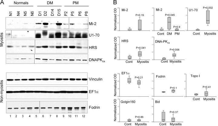
Unique autoantibody specificities are strongly associated with distinct clinical phenotypes, making autoantibodies useful for diagnosis and prognosis. To investigate the mechanisms underlying this striking association, we examined autoantigen expression in normal muscle and in muscle from patients with autoimmune myositis. Although myositis autoantigens are expressed at very low levels in control muscle, they are found at high levels in myositis muscle. Furthermore, increased autoantigen expression correlates with differentiation state, such that myositis autoantigen expression is increased in cells that have features of regenerating muscle cells. Consistent with this, we found that cultured myoblasts express high levels of autoantigens, which are strikingly down-regulated as cells differentiate into myotubes in vitro. These data strongly implicate regenerating muscle cells rather than mature myotubes as the source of ongoing antigen supply in autoimmune myositis. Myositis autoantigen expression is also markedly increased in several cancers known to be associated with autoimmune myositis, but not in their related normal tissues, demonstrating that tumor cells and undifferentiated myoblasts are antigenically similar. We propose that in cancer-associated myositis, an autoimmune response directed against cancer cross-reacts with regenerating muscle cells, enabling a feed-forward loop of tissue damage and antigen selection. Regulating pathways of antigen expression may provide unrecognized therapeutic opportunities in autoimmune diseases.


Figures

Figure 1.
Figure 1.
Myositis-specific autoantigens are expressed at low levels in control muscle and at high levels in myositis muscle. (A) Equal protein amounts of lysed human muscle biopsies, obtained from normal, DM, and PM patients, were immunoblotted with antibodies against the indicated antigens. The myositis-specific autoantigens (Mi-2, U1-70 kD, HRS, and DNA-PKcs) were expressed at high levels in myositis muscle. In contrast, levels of nonmyositis autoantigens and nonautoantigens were not increased in the myositis tissue. Of note, immunoblotted U1-70 kD was detected in two different forms in the tissue lysates (70 and 68 kD). These represent different phosphorylation states of U1-70 kD. Such altered forms can be detected in vitro; U1-70 kD in unincubated cell lysates migrates at 70 kD, but after incubating the lysates at 37°C for 30 min with λ-phosphatase, only the 68-kD form is blotted (not depicted). Black lines indicate that intervening lanes have been spliced out. (B) The immunoblots were scanned, and the data were quantitated by normalizing the immunoblotted level of each autoantigen relative to that of vinculin in the same lysate. In the set of immunoblots shown (representative of two to six blots performed with each different lysed muscle biopsy), the vinculin data shown in the fifth panel was used to normalize all of the immunoblotted antigens except fodrin. The α-fodrin data were obtained using a different set of lysates, with its own matched vinculin blot (the latter is not depicted), and these vinculin values were used to normalize the α-fodrin data.
Figure 2.
Figure 2.
DNA-PKcs, HRS, and Ku70 staining is increased in myocytes in DM and PM muscle compared with normal muscle. (A–C) Normal (A) and PM (B and C) muscle biopsies were stained using affinity-purified rabbit anti-HRS polyclonal antibody in the absence (A and B) or presence (C) of competing peptide. A faint sarcoplasmic pattern was noted in normal muscle (A), with increased staining being detected in the PM biopsy (B). The staining was abolished upon preincubation of the antibody with specific (immunizing) peptide (C), but not with a nonspecific peptide (not depicted). (D–I) Normal (D and G), PM (E and H), and DM (F and I) biopsies were stained using monoclonal antibodies against DNA-PKcs (D–F) or Ku70 (G–I). Normal biopsies showed low levels of nuclear staining. The number and intensity of brown positive nuclei was significantly increased in DM and PM biopsies. All panels have a magnification of 40. The data shown are representative fields from multiple different normals and patients. A, n = 5; B and C, n = 3; D, n = 4; E, n = 12; and F, n = 4.
Figure 3.
Figure 3.
NCAM and neonatal MHC-positive muscle fibers are rare in controls, but frequent in muscle biopsies from DM and PM patients. (A–F) Normal (A and D), PM (B and E), and DM (C and F) biopsies were stained using antibodies against NCAM (A–C) or neonatal myosin heavy chain (nMHC) (D–F). NCAM and nMHC-positive fibers were rarely detected in normal muscle, but were frequently seen in PM and DM biopsies. Positive fibers were enriched in perifascicular areas of DM (C and F) and were spread throughout the fascicle in PM (B and E). The data shown are representative fields from multiple different normals and patients; normal, n = 5; DM, n = 11; and PM, n = 11. All fields have a magnification of 40. (G) To quantitate the data, the number of NCAM-positive fibers in up to five different areas of the biopsy were counted, and the average number of positive fibers per field was calculated. Normal biopsies occasionally showed a small number of NCAM-positive fibers (not depicted in A). (H) Equal protein amounts of muscle biopsy lysates obtained from normal, DM, and PM patients were immunoblotted with antibody against NCAM. Black lines indicate that intervening lanes have been spliced out.
Figure 4.
Figure 4.
High levels of DNA-PKcs and HRS staining are found in regenerating myocytes in DM muscle. (A) Serial DM muscle biopsy sections were stained with antibodies against neonatal myosin heavy chain or DNA-PKcs. (left and center) Both antibodies stained an overlapping subset of cells in serial sections with a striking perifascicular pattern characteristic of DM. Note that areas within the fascicle are not stained with either antibody. (right) Higher magnification showed increased DNA-PKcs staining in many muscle cell nuclei. (B) A separate set of serial DM muscle biopsy sections was stained with antibodies against NCAM (monoclonal) or HRS (affinity-purified anti-HRS polyclonal antibody). These antibodies stained an overlapping subset of cells on the serial sections. (A and B) Unless otherwise indicated, all panels have a magnification of 5.
Figure 5.
Figure 5.
Autoantigen expression is high in cultured myoblasts and low in differentiated myotubes. Primary human myoblasts were differentiated to myotubes over 14 d as described in Materials and methods. The levels of expressed antigens were assessed by immunoblotting equal protein amounts of the lysates harvested at each time point. (A) Lysates made from cells harvested at day 0 (myoblasts) and day 14 of differentiation (myotubes) were immunoblotted with antibodies against NCAM and myosin. Expression of NCAM and myosin in myoblasts and myotubes, respectively, confirmed the differentiation status of the cell cultures. (B) Lysates made from cells harvested at various times during the 14 d differentiation period were immunoblotted with the indicated antibodies. Levels of autoantigen expression were decreased in myotubes; in contrast, the expression of nonautoantigens remained constant. (C) The data obtained in B was scanned and quantitated. For each antigen, the myoblast level (day 0 of differentiation) was assigned the value 100%, and the antigen levels at each time point were expressed relative to this.
Figure 6.
Figure 6.
Myositis-specific autoantigen levels are not increased in proliferating versus quiescent aortic smooth muscle cells. Proliferating (P) or quiescent (Q) human aortic smooth muscle cells were prepared as described Materials and methods. 3 × 10⁴ cells were electrophoresed in each gel lane. Lysates were immunoblotted with the indicated antibodies. The data were scanned, and the levels of each antigen in the proliferating cultures was expressed relative to that in the quiescent culture (P/Q ratio). PCNA and α-smooth muscle actin were expressed at high levels in the proliferating and quiescent cultures, respectively. DNA-PKcs and Mi-2 expression were not significantly different in proliferating and quiescent cells.
Figure 7.
Figure 7.
Myositis autoantigens are frequently expressed at low levels in normal tissue, but at increased levels in tumors. (A and B) Lysates were made from the indicated normal and tumorous human tissues. Equal protein amounts were electrophoresed in each gel lane on 10% SDS-polyacrylamide gels, and immunoblotted with a human serum monospecific for Mi-2, a polyclonal anti-HRS antibody, or monoclonal antibodies against DNA-PKcs and vinculin. The data shown are representative of those obtained from 8–10 different cancer biopsies of each type in two to three separate experiments. Black lines indicate that intervening lanes have been spliced out. (B) Mi-2 levels were elevated in 10/10 (breast) and 9/10 (lung) cancer tissue lysates; DNA-PKcs expression was increased in 9/10 (breast) and 8/8 (lung) cancer tissue lysates, and HRS levels were elevated in 3/3 (breast) cancer tissue lysates. HRS was expressed fairly robustly in normal lung with only small increases (if any) detected in cancerous lung.

References

    1. von Muhlen, C.A., and E.M. Tan. 1995. Autoantibodies in the diagnosis of systemic rheumatic diseases. Semin. Arthitis Rheum. 24:323–358. - PubMed
    1. Plotz, P.H. 2003. The autoantibody repertoire: searching for order. Nat. Rev. Immunol. 3:73–78. - PubMed
    1. Targoff, I.N., and M. Reichlin. 1985. The association between Mi-2 antibodies and dermatomyositis. Arthritis Rheum. 28:796–803. - PubMed
    1. Love, L.A., R.L. Leff, D.D. Fraser, I.N. Targoff, M. Dalakas, P.H. Plotz, and F.W. Miller. 1991. A new approach to the classification of idiopathic inflammatory myopathy: myositis-specific autoantibodies define useful homogeneous patient groups. Medicine (Baltimore). 70:360–374. - PubMed
    1. Arnett, F.C., T.J. Hirsch, W.B. Bias, M. Nishikai, and M. Reichlin. 1981. The Jo-1 antibody system in myositis: relationships to clinical features and HLA. J. Rheumatol. 8:925–930. - PubMed

Publication types