. 2005 Mar 1;102(9):3254-9.
doi: 10.1073/pnas.0500327102. Epub 2005 Feb 22.

Assembly of the bacteriophage T4 primosome: single-molecule and ensemble studies

Affiliations

Assembly of the bacteriophage T4 primosome: single-molecule and ensemble studies

Zhiquan Zhang et al. Proc Natl Acad Sci U S A. .

Erratum in

  • Proc Natl Acad Sci U S A. 2005 Sep 13;102(37):13349-50

Abstract

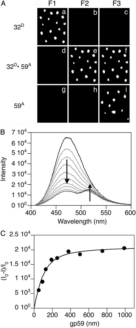
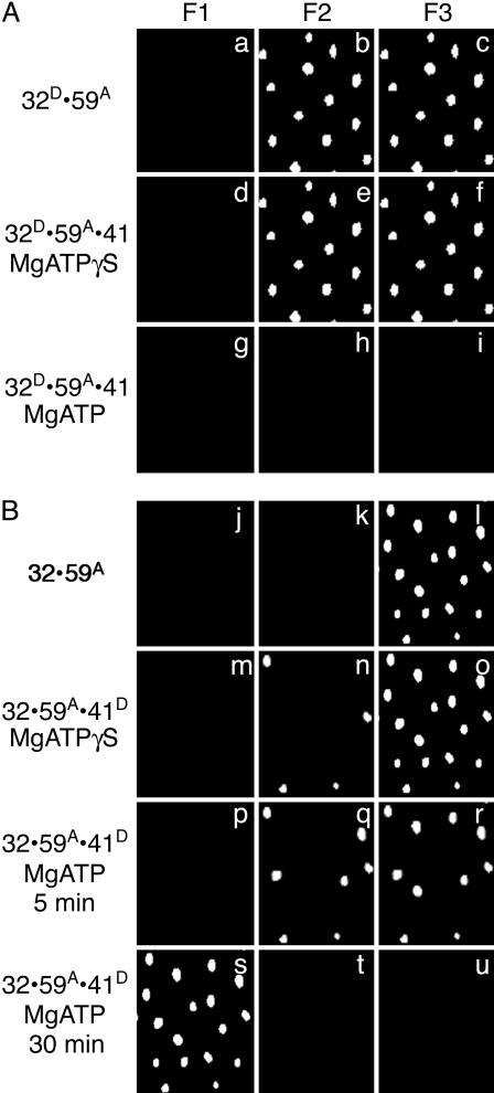
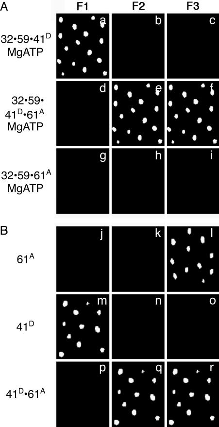
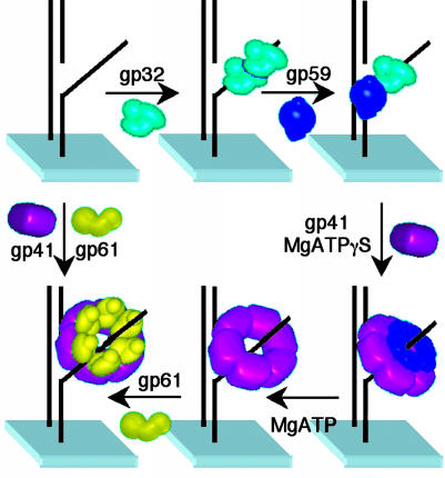
Within replisomes for DNA replication, the primosome is responsible for unwinding double-stranded DNA and synthesizing RNA primers. Assembly of the bacteriophage T4 primosome on individual molecules of ssDNA or forked DNA (fDNA) has been studied by using FRET microscopy. On either DNA substrate, an ordered process of assembly begins with tight 1:1 binding of ssDNA-binding protein (gp32) and helicase-loading protein (gp59) to the DNA. Magnesium adenosine 5'-O-(3-thiotriphosphate) (MgATPgammaS) mediates the weak binding of helicase (gp41) to DNA coated with gp32 and gp59, whereas MgATP induces gp32 and gp59 to dissociate, leaving gp41 bound to the DNA. Finally, primase (gp61) binds to the gp41.DNA complex. Ensemble studies were used to determine protein stoichiometries and binding constants. These single-molecule studies provide an unambiguous description of the pathway for assembly of the primosome on the lagging strand of DNA at a replication fork.


Figures

Fig. 1.
Fig. 1.
Protein–protein interactions between gp32 and gp59 on fDNA. (A) The fluorescence from individual molecules of fDNA with the proteins bound in the order as indicated at the side of each row. The gp32 protein is labeled with A488 (gp32D) and the gp59 protein is labeled with A555 (gp59A). The filter sets are described in Experimental Methods: F1 is for A488 emission, F2 for FRET between A488 and A555, and F3 for A555 emission. (B) Ensemble FRET studies of Oregon-green-488-maleimide-labeled gp59 titrated into a solution of 400 nM CPM-labeled gp32 and 100 nM fDNA. The fluorescence spectra of 400 nM CPM-gp32 alone (black line), the endpoint of the titration at 1 μM Oregon-green-488-maleimide-gp59 (dark gray line), and several intermediate spectra (light gray lines) are shown. (C) Analysis of the donor quenching and acceptor sensitization plotted against the gp59 concentration determines the stoichiometry among gp32, gp59, and fDNA to be 1:1:1 with a calculated binding constant of ≈40 nM.
Fig. 2.
Fig. 2.
The ATP-dependent loading of gp41 by gp59 onto fDNA coated with gp32. The fluorescence from individual molecules of fDNA with the proteins bound in the order as indicated at the side of each row. (A) The gp32 protein is labeled with A488 (gp32D), the gp59 protein is labeled with A555 (gp59A), and the gp41 protein is unlabeled. MgATPγS (500 μM) is present for the sample in row 2, and 500 μM MgATP is present for the sample in row 3. (B) The gp32 protein is unlabeled, the gp59 protein is labeled with A555 (gp59A), and the gp41 is labeled with A488 (gp41D). MgATPγS (500 μM) is present for the sample in row 2, and 500 μM MgATP is present for the sample in row 3 (5 min after addition of gp41 and nucleotide) and in row 4 (30 min after addition of gp41 and nucleotide). The filter sets are described in the legend to Fig. 1.
Fig. 3.
Fig. 3.
Completed primosome formation by the addition of gp61 or an alternative, direct formation of the gp41/gp61 primosome. The fluorescence from individual molecules of fDNA with the proteins bound in the order as indicated at the side of each row. (A) The gp32 and gp59 proteins are unlabeled, the gp41 protein is labeled with A488 (gp41D), and the gp61 protein is labeled with A555 (gp61A) in the presence of 500 μM MgATP. (B) The gp41 protein is labeled with A488 (gp41D), and the gp61 protein is labeled with A555 (gp61A) in the absence of MgATP. The filter sets are described in the legend to Fig. 1.
Fig. 4.
Fig. 4.
Assembly mechanism of the T4 lagging-strand primosome on fDNA. The gp32 protein binds to fDNA with either subsequent or concurrent binding of gp59. Subsequently, gp41 binds to gp59 and is loaded onto the fDNA in the presence of nucleotide. ATP hydrolysis is required for gp41 to displace gp32 and gp59, either directly or by translocation. The gp61 protein then binds and interacts closely with gp41 on fDNA. In the absence of gp32 and gp59, both gp41 and gp61 bind to fDNA.

References

    1. Benkovic, S. J., Valentine, A. M. & Salinas, F. (2001) Annu. Rev. Biochem. 70, 181-208. - PubMed
    1. Yu, X., Jexewska, M. J., Bujalowski, W. & Egelman, E. H. (1996) J. Mol. Biol. 259, 7-14. - PubMed
    1. Keck, J. L. & Berger, J. M. (2001) Nat. Struct. Biol. 8, 2-4. - PubMed
    1. Baker, T. A. & Bell, S. P. (1998) Cell 92, 295-305. - PubMed
    1. Liu, C. C. & Alberts, B. M. (1981) J. Biol. Chem. 256, 2813-2820. - PubMed

Publication types

MeSH terms

LinkOut - more resources