. 2005 Mar 1;102(9):3248-53.
doi: 10.1073/pnas.0409851102. Epub 2005 Feb 22.

Crystal structure of DNA sequence specificity subunit of a type I restriction-modification enzyme and its functional implications

Affiliations

Crystal structure of DNA sequence specificity subunit of a type I restriction-modification enzyme and its functional implications

Jeong-Sun Kim et al. Proc Natl Acad Sci U S A. .

Abstract

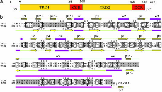
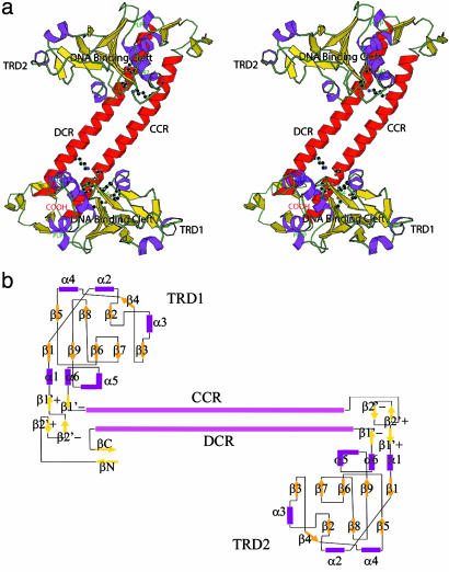
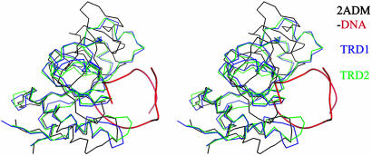
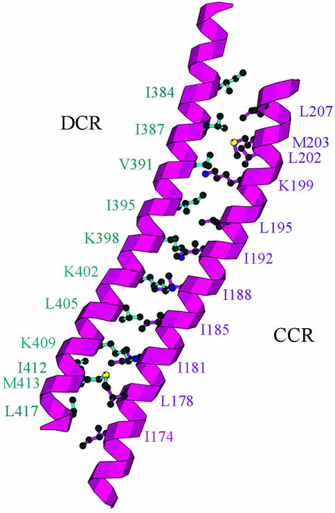
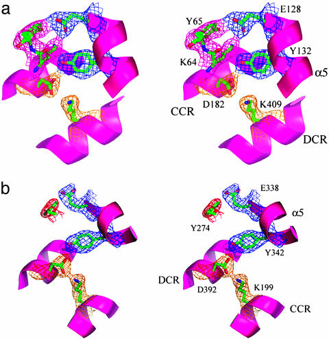
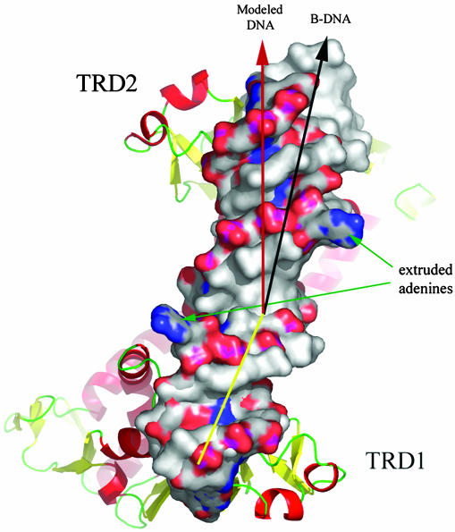
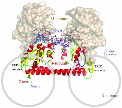
Type I restriction-modification enzymes are differentiated from type II and type III enzymes by their recognition of two specific dsDNA sequences separated by a given spacer and cleaving DNA randomly away from the recognition sites. They are oligomeric proteins formed by three subunits: a specificity subunit, a methylation subunit, and a restriction subunit. We solved the crystal structure of a specificity subunit from Methanococcus jannaschii at 2.4-A resolution. Two highly conserved regions (CRs) in the middle and at the C terminus form a coiled-coil of long antiparallel alpha-helices. Two target recognition domains form globular structures with almost identical topologies and two separate DNA binding clefts with a modeled DNA helix axis positioned across the CR helices. The structure suggests that the coiled-coil CRs act as a molecular ruler for the separation between two recognized DNA sequences. Furthermore, the relative orientation of the two DNA binding clefts suggests kinking of bound dsDNA and exposing of target adenines from the recognized DNA sequences.


Figures

Fig. 1.
Fig. 1.
Domain assignment and secondary structure of S-subunit of a type I R-M from M. jannschii. (a) Schematic representation of an S-subunit. (b) Sequence alignment of TRD1/TRD2 and CCR/DCR. Identical residues have a gray background, and highly homologous and low homologous residues are in small and larger boxes, respectively.
Fig. 2.
Fig. 2.
Tertiary structure representation of an S-subunit. (a) Stereogram of the overall structure. The monomeric structure is displayed by ribbon representation and hydrogen-bonding residues at domain interfaces are indicated by stick models. The highly conserved PLPP sequences and DNA binding clefts are marked. A monomer consists of four successive structural domains: the globular TRD1, a long helical CCR domain, the globular TRD2, and a C-terminal DCR helix. Two almost identical TRDs are attached to both ends of complementary CR helices. The N and C termini form a β-strand and a β-sheet to close both termini. The figure was prepared by using molscript (35). (b) Topology diagram of the S-subunit of a type I R-M system from M. jannaschii. Two TRDs are symmetrically located around the complementary CR helices, except for the two β-strands (βN and βC) at the N and C terminus of TRD1.
Fig. 3.
Fig. 3.
Stereogram of superposed TRD1 and TRD2 on the DNA binding domain of the TaqI-MTase. TRD1, TRD2, and TaqI-MTase are differentiated by colors, and the bound DNA backbone to the DNA binding domain of TaqI-MTase is displayed in red. The three globular structures superpose well especially in the DNA binding region.
Fig. 4.
Fig. 4.
Coiled–coil structures of CRs. Two highly CRs in the middle and at the end of the primary sequences of an S-subunit form long coiled–coil structures with a large number of hydrophobic residues between the two helical coils.
Fig. 5.
Fig. 5.
Stereo presentations of the TRD's interaction with the CRs. The longest helix, α5, in the TRDs interacts with CR helices by providing hydrophobic residues at the ends of the CRs as well as hydrophilic residues toward the middle of CR helices that were displayed with 2 Fo - Fc density contoured at 1.5 σ. The figures were prepared by using pymol (www.pymol.org). (a) Lys-64 in TRD1 is located at the interface with the CRs and has a well defined electron density. However, it has a distorted geometry. (b) The interaction of TRD2 with CRs is simplified compared with that of TRD1 probably because of the loss of a hydrogen bond donor corresponding to Lys-64 in TRD1.
Fig. 6.
Fig. 6.
Model for DNA binding to the S-subunit of an M. janaschii type I R-M system. The model was obtained by transporting the DNA from the TaqI-MTase DNA complex structure to each TRD of the S-subunit after the DNA binding domain of TaqI-MTase was superposed to TRDs of the S-subunit. TRDs interact with the DNA major groove. When the bound DNA in a colored surface presentation is compared with normal B-DNA in a gray surface presentation, the modeled DNA is kinked and twisted toward one end of the DNA.
Fig. 7.
Fig. 7.
Model for subunit assembly. The S-subunit is drawn as a ribbon diagram, and the obtained DNA model shown in sticks. The M-subunit of the TaqI-MTase is drawn as a surface model and was docked around the TRDs of the S-subunit, based on the sequence similarity and functional features. The possible R-subunit binding regions drawn as gray circles are marked based on the structural features as well as the reported experimental data.

References

    1. Studier, F. W. & Bandyopadhyay, P. K. (1998) Proc. Natl. Acad. Sci. USA 85, 4677-4681. - PMC - PubMed
    1. Dreier, J., MacWilliams, M. P. & Bickle, T. A. (1996) J. Mol. Biol. 264, 722-733. - PubMed
    1. Bickle, T. A. & Krüger, D. H. (1993) Microbiol. Rev. 57, 434-450. - PMC - PubMed
    1. Redaschi, N. & Bickle, T. A. (1996) J. Mol. Biol. 257, 790-803. - PubMed
    1. Yuan, R. (1981) Annu. Rev. Biochem. 50, 285-315. - PubMed

Publication types

Associated data