. 2005 Mar 8;102(10):3685-90.
doi: 10.1073/pnas.0408217102. Epub 2005 Feb 22.

Matrix metalloproteinase 13 mediates nitric oxide activation of endothelial cell migration

Affiliations

Matrix metalloproteinase 13 mediates nitric oxide activation of endothelial cell migration

Esther López-Rivera et al. Proc Natl Acad Sci U S A. .

Abstract

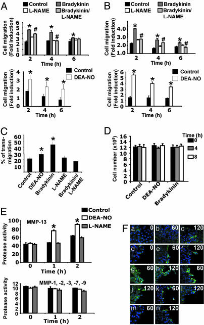
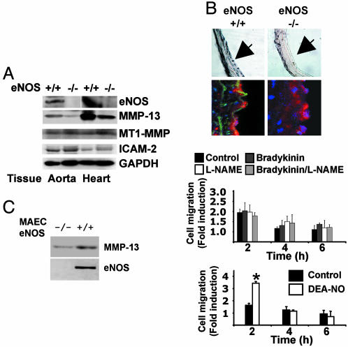
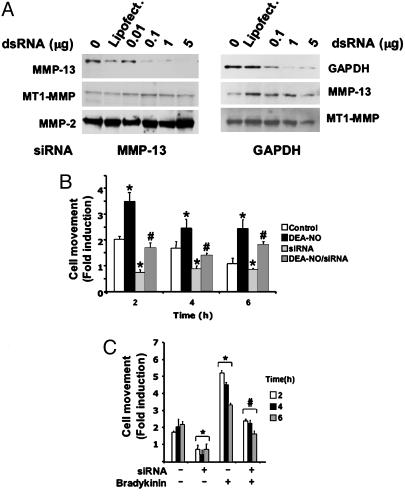
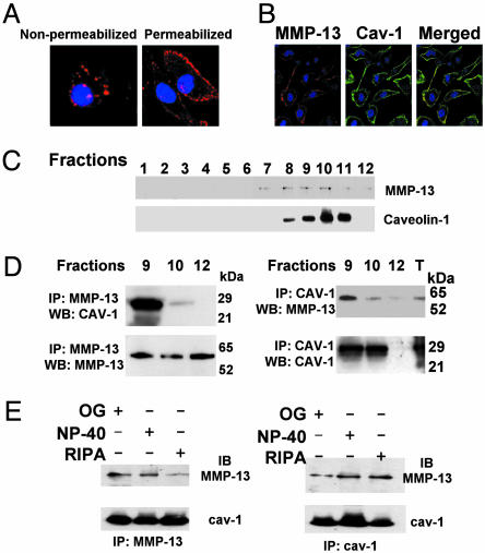
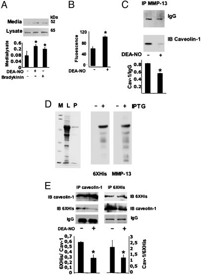
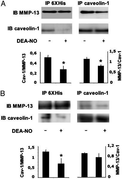
To explore the mechanisms by which NO elicits endothelial cell (EC) migration we used murine and bovine aortic ECs in an in vitro wound-healing model. We found that exogenous or endogenous NO stimulated EC migration. Moreover, migration was significantly delayed in ECs derived from endothelial NO synthase-deficient mice compared with WT murine aortic EC. To assess the contribution of matrix metalloproteinase (MMP)-13 to NO-mediated EC migration, we used RNA interference to silence MMP-13 expression in ECs. Migration was delayed in cells in which MMP-13 was silenced. In untreated cells MMP-13 was localized to caveolae, forming a complex with caveolin-1. Stimulation with NO disrupted this complex and significantly increased extracellular MMP-13 abundance, leading to collagen breakdown. Our findings show that MMP-13 is an important effector of NO-activated endothelial migration.


Figures

Fig. 1.
Fig. 1.
NO induces EC movement in a wound-healing model. (A and B) BAEC (A) or MAEC (B) monolayers were injured and treated with 10-6 M bradykinin, 500 μM l-NAME, bradykinin/l-NAME, or 100 μM DEA-NO, and cell movement was monitored by microscopy (n = 3 by triplicate; mean ± SD; *, P < 0.05 vs. control; #, P < 0.05 vs. bradykinin). (C) BAEC monolayers were seeded on 8-μm porous transwell filters and treated with 100 μM DEA-NO, 10-6 M bradykinin, 0.5 mM l-NAME, or the combination bradykinin/l-NAME. After 6 h of plating, transmigration was evaluated by confocal microscopy (see Methods for details, n = 2 by triplicate; *, P < 0.05 vs. control). (D) ECs were injured and treated with 100 μM DEA-NO or 10-6 M bradykinin. Total cell number was evaluated at the indicated times (n = 3 by triplicate; mean ± SD). (E) EC monolayers were treated with 100 μM DEA-NO or 500 μM l-NAME. MMP activities were measured by fluorimetry from culture supernatants collected at regular time points (n = 3 by quadruplicate; mean ± SD; *, P < 0.05 vs. control). (F) EC monolayers (ac) were injured (dn, see Methods for details) and treated with 100 μM DEA-NO (b, c, g, and h), 100 μM DEA (m and n), 10-6 M bradykinin (i and j), or 10-6 M bradykinin/500 μM l-NAME (k and l). MMP-13 was visualized over time (0, 1, and 2 has indicated) with immunohistochemical staining (MMP-13, FITC, green; nuclei Hoechst, blue) by confocal microscopy (magnification: ×40, n = 3).
Fig. 2.
Fig. 2.
ECs from eNOS-deficient mice migrate slower and show lower MMP-13 levels when compared with their eNOS WT counterparts. (A) Aortas and hearts from a pool of eNOS-deficient mice and eNOS WT mice were homogenized and used to evaluate eNOS, MMP-13, MT1-MMP, ICAM-2, and GAPDH expression (n = 3 animals by triplicate). (B) Aortic rings were isolated from eNOS-deficient mice and eNOS WT mice, and MMP-13 was visualized by immunohistochemistry and immunohistofluorescence using an anti-MMP-13 antibody. MMP-13 was visualized by peroxidase staining (Upper, magnification ×20) and FITC (Lower, magnification ×60). Aortic nuclei were stained with Hoechst. ICAM-2 expression was visualized in red (n = 5). (C) MAECs from eNOS WT or eNOS-deficient mice were isolated, and lysates were immunobloted to detect the expression of MMP-13 and eNOS (Left). In addition, a wound-healing assay was performed in cells treated with 100 μM DEA-NO, 10-6 M bradykinin, 500 μM l-NAME, and bradykinin/l-NAME (n = 3 by triplicate; mean ± SD; *, P < 0.01 vs. control).
Fig. 3.
Fig. 3.
EC migration depends on MMP-13 expression. (A) Immunoblot detection of MMP-13, MT1-MMP, and MMP-2 (Left) and GAPDH, MMP-13, and MT1-MMP (Right) in MMP-13 (Left) and GAPDH (Right) silenced cells. Shown is one representative experiment from a total of three. (B) ECs were injured and treated with 100 μM DEA-NO (n = 3 by quadruplicate; mean ± SD; *, P < 0.05 vs. control; #, P < 0.05 vs. DEA-NO). (C) ECs were subject or not to MMP-13 silencing, injured, and treated with 10-6 M bradykinin (n = 3 by triplicate; mean ± SD; *, P < 0.05 vs. nonstimulated/nonsilenced; #, P < 0.05 vs. stimulated with bradykinin/nonsilenced).
Fig. 4.
Fig. 4.
MMP-13 colocalizes with caveolin-1 in endothelial plasma membranes. (A) Confocal microscopy analysis of BAECs showing the expression of MMP-13 in nonpermeabilized and permeabilized BAECs. (B) Confocal microscopy analysis showing the expression of caveolin-1 (green) and MMP-13 (red) in BAECs. Merged panel shows colocalization of MMP-13 and caveolin-1 (yellow) (n = 5). (C) Caveolae-enriched fractions were isolated and identified by immunoblot with anti-caveolin-1. MMP-13 was also evaluated in the same fractions (n = 3). (D) Caveolin-1-positive (fractions 9 and 10) and -negative (fraction 12) fractions from ECs were subjected to cross-coimmunoprecipitation with anti-MMP-13 (Left) and anti-caveolin-1 (Right). Caveolin-1 or MMP-13 were detected by immunoblot (n = 4). (E) Cell lysates were subjected to cross-coimmunoprecipitation as indicated, in octyl-glucoside (OG), Nonidet P-40, and RIPA buffers (n = 3).
Fig. 5.
Fig. 5.
NO disrupts the MMP-13/caveolin-1 complex in BAEC. (A) Immunoblot of MMP-13 from cell lysates or culture media collected after 2 h of treatment of ECs with 100 μM DEA-NO or 10-6 M bradykinin. The graph represents the densitometric analysis of data from three independent experiments (mean ± SD; *, P < 0.05). (B) MMP-13 activity assay in cell media from ECs treated for 2 h with 100 μM DEA-NO (n = 3; mean ± SD; P < 0.05). (C) Immunoprecipitation of MMP-13 from caveolin-1-enriched fractions of vehicle and ECs treated with 100 μM DEA-NO. Caveolin-1 was detected by immunoblot (n = 4 by triplicate; mean ± SD; *, P < 0.01). (D) MMP-13 was expressed in E. coli and purified by affinity chromatography. IP, immunoprecipitation; WB, Western blot. (Left) Coomassie staining of a 12% SDS/PAGE. M, molecular weight marker; L, bacterial lysate from cells treated with 1 mM isopropyl β-d-thiogalactoside (IPTG); P, purified MMP-13. (Right) Immunoblots from purified MMP-13, with anti-6XHis and anti-MMP-13 antibodies. (E) Crossed-coimmunoprecipitation experiments from purified MMP-13 incubated with cell lysates of BAECs for 16 h at 4°C and treated with DEA-NO or vehicle for 2 h (n = 3; mean ± SD; *, P < 0.05).
Fig. 6.
Fig. 6.
NO disrupts the in vitro MMP-13/caveolin-1 complex. (A) Recombinant 6XHis–MMP-13 and GST–caveolin-1 were purified and subject to immunoprecipitation (IP) experiments in the presence or absence of 100 μM DEA-NO to visualize the binding to caveolin-1 and MMP-13, respectively (n = 3; mean ± SD; *, P < 0.05). IB, immunoblot. (B) Recombinant 6XHis–MMP-13 or GST–caveolin-1 were pretreated with 100 μM DEA-NO for 1 h, bound to GST–caveolin or 6XHis–MMP-13, respectively (nontreated with DEA-NO), and subjected to immunoprecipitation (IP) experiments (n = 3; mean ± SD; *, P < 0.05). IB, immunoblot.

Similar articles

Cited by

References

    1. Schwentker, A., Vodovotz, Y., Weller, R. & Billiar, T. R. (2002) Nitric Oxide 7, 1-10. - PubMed
    1. Papapetropoulos, A., Garcia-Cardena, G., Madri, J. A. & Sessa, W. C. (1997) J. Clin. Invest. 100, 3131-3139. - PMC - PubMed
    1. Ziche, M., Morbidelli, L., Choudhuri, R., Zhang, H. T., Donnini, S., Granger, H. J. & Bicknell, R. (1997) J. Clin. Invest. 99, 2625-2634. - PMC - PubMed
    1. Dimmeler, S., Dernbach, E. & Zeiher, A. M. (2000) FEBS Lett. 477, 258-262. - PubMed
    1. Noiri, E., Lee, E., Testa, J., Quigley, J., Colflesh, D., Keese, C. R., Giaever, I. & Goligorsky, M. S. (1998) Am. J. Physiol. 274, C236-C244. - PubMed

Publication types

LinkOut - more resources