. 2005 Mar 1;174(5):2591-601.
doi: 10.4049/jimmunol.174.5.2591.

CD8+ T cell immunity against a tumor/self-antigen is augmented by CD4+ T helper cells and hindered by naturally occurring T regulatory cells

Affiliations

CD8+ T cell immunity against a tumor/self-antigen is augmented by CD4+ T helper cells and hindered by naturally occurring T regulatory cells

Paul A Antony et al. J Immunol. .

Abstract

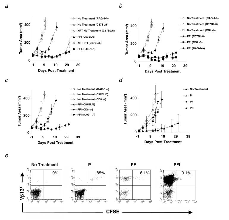
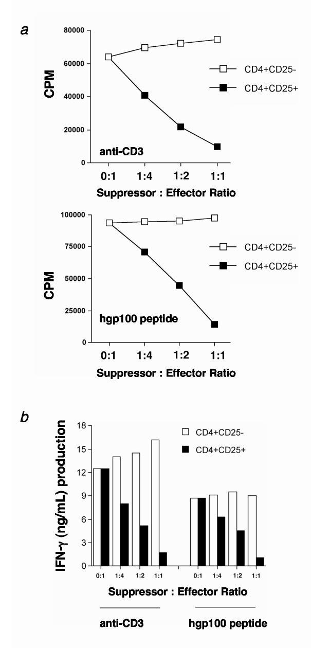
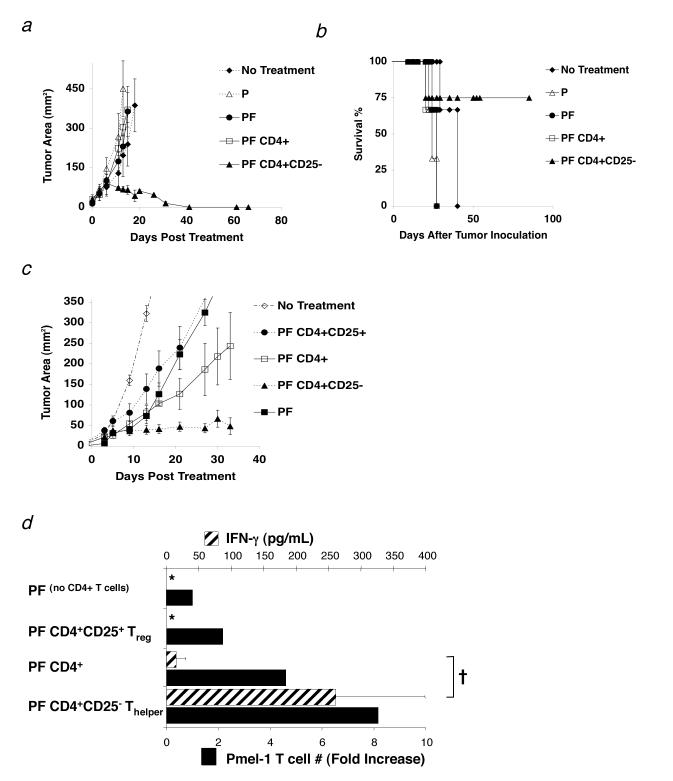
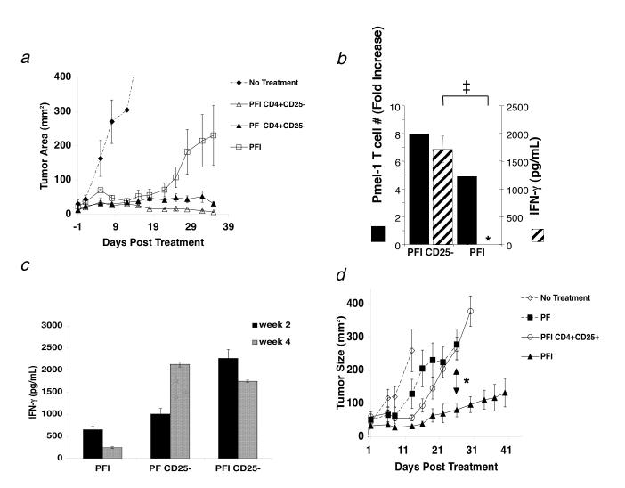
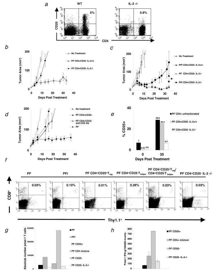
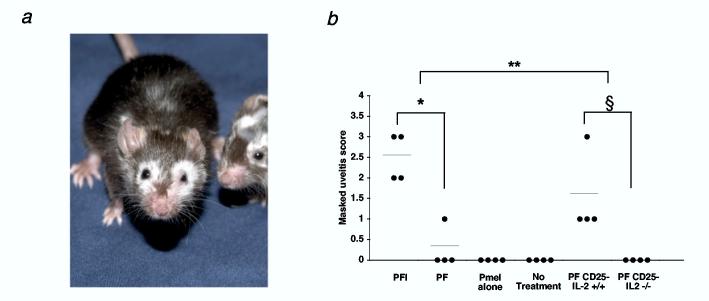
CD4(+) T cells control the effector function, memory, and maintenance of CD8(+) T cells. Paradoxically, we found that absence of CD4(+) T cells enhanced adoptive immunotherapy of cancer when using CD8(+) T cells directed against a persisting tumor/self-Ag. However, adoptive transfer of CD4(+)CD25(-) Th cells (Th cells) with tumor/self-reactive CD8(+) T cells and vaccination into CD4(+) T cell-deficient hosts induced autoimmunity and regression of established melanoma. Transfer of CD4(+) T cells that contained a mixture of Th and CD4(+)CD25(+) T regulatory cells (T(reg) cells) or T(reg) cells alone prevented effective adoptive immunotherapy. Maintenance of CD8(+) T cell numbers and function was dependent on Th cells that were capable of IL-2 production because therapy failed when Th cells were derived from IL-2(-/-) mice. These findings reveal that Th cells can help break tolerance to a persisting self-Ag and treat established tumors through an IL-2-dependent mechanism, but requires simultaneous absence of naturally occurring T(reg) cells to be effective.


Figures

Figure 1
Figure 1
Naturally occurring CD4+ T cells inhibit effective immunotherapy to established tumors. (a-d) Mice were inoculated with 1.0 × 10⁵ cells of B16 melanoma on day -14 before adoptive cell transfer with 1.0 × 10⁶ pmel-1 T cells (P), 2.0 × 10⁷ PFU rFPVhgp100 (F), and 600,000 IU of exogenous IL-2 (I), which was given daily for 3-4 days. (a) Tumor regression in C57BL/6 mice (□) is compared with RAG-1-/- mice (•) and C57BL/6 mice receiving 500 (cGy) whole body irradiation on day -1 of treatment (□). (b) Tumor regression in C57BL/6 mice (□) is compared with RAG-1-/- mice (•) and CD4-/- mice (□). (c) Tumor regression in C57BL/6 mice (□) is compared with RAG-1-/- mice (•) and CD8-/- mice (◆) in the same experiment. Data are represented as mean tumor size ± S.E.M. Experiments were independently repeated twice. (d-e) Tumor regression is IL-2 dependent. (d) Transfer of pmel-1 T cells alone (Δ) or pmel-1 T cells and rFPVhgp100 vaccine (•) into tumor-bearing RAG-1-/- hosts is similar to no treatment (◆). Addition of exogenous IL-2 with cells and vaccine is required for tumor regression (□). (e) CFSE profile of adoptively transferred pmel-1 T cells into RAG-1-/- hosts. Pmel-1 CD8+ T cells were labeled with CFSE and adoptively transferred into tumor-bearing RAG-1-/- hosts alone (P), with vaccination (PF), or with vaccination and exogenous IL-2 (PFI). 4 days later, splenocytes from treated mice were analyzed by flow cytometry. Gated on CD8+ T cells and displayed as Vβ13-PE vs. CFSE.
Figure 2
Figure 2
CD4+CD25+ Treg cells suppress pmel-1 CD8+ T cell proliferation and function in vitro. (a-b) Highly purified Treg cells from C57BL/6 lymph nodes suppress (a) proliferation and (b) IFN-γ production of pmel-1 T cells in vitro. Treg or Thelper cells were activated with irradiated APC (1:1 ratio) and soluble anti-CD3 antibody (0.5 μg/mL) for 3 days in the presence of 100 IU human IL-2. Cells were then split and maintained for an additional 7-14 days in 100 IU human IL-2. (a) Pmel-1 T cells were stimulated with soluble anti-CD3 or 1 μM hgp10025-33 peptide-pulsed T-depleted spleen cells in the presence of activated Treg (■) or Thelper (□) cells for 3 days in the absence of IL-2 at the indicated ratios. Maximum suppression of pmel-1 T cell proliferation is seen at a 1:1 suppressor to effector ratio. (b) Pmel-1 T cells were stimulated with anti-CD3 or 1 μM hgp10025-33 peptide-pulsed TΔS cells in the presence of activated Treg (■) or Thelper (□) cells for 3 days in the absence of IL-2 at the indicated ratios. Maximal suppression of IFN-γ production by pmel-1 T cells for both modes of stimulation is seen at a 1:1 suppressor to effector ratio. Experiments were independently repeated twice.
Figure 3
Figure 3
Thelper cells maintain self-reactive effector CD8+ T cells in vivo. (a-d) RAG-1-/-mice were inoculated with 3.0 × 10⁵ cells of B16 melanoma between day -7 and -14 before adoptive cell transfer. (a) Thelper cells help CD8+ T cell mediated antitumor immunity to established B16 melanoma. Mice receiving 1.0 × 10⁶ pmel-1 T cells (P), 2.0 × 10⁷ PFU fowlpox virus encoding human gp100 (F), and 1.0 × 10⁶ Thelper cells maintained long-term, durable regression of established B16 melanoma (□). Data are represented as mean tumor size ± S.E.M. Data represent 6 independent experiments. (b) Survival of mice in (a) treated with pmel-1 T cells (P), rFPVhgp100 vaccine (F), and Thelper cells was maintained up to 80 days post treatment (□). (c) Adoptive cell transfer of sorted 1.0 × 10⁵ Treg cells (•) or a mixture of 1.0 × 10⁵ Treg and 1.0 × 10⁶ Thelper cells (1:10 ratio; CD4+ T cells; □) with pmel-1 T cells and vaccination does not maintain tumor regression of B16 melanoma. Removal of the Treg subset from the unfractionated CD4+ T cell pool allows the helper function of the remaining Thelper cells to become apparent (□). Data represent 5 independent experiments. Experiment in (c) was stopped at 35 days post treatment to allow for analysis of adoptively transferred pmel-1 T cells. (d) Thelper cells maintain maximal pmel-1 T cell numbers (solid black bars) and function (cross-hatched bars) in the absence of IL-2. †, P <0.037. Proliferation bar graphs represent fold increase in the absolute number of pmel-1 T cells (% CD8+ Thy1.1+ Vβ13+ T cells × splenocyte count) taken from pooled spleens 5 wks after transfer (n=2). Fold increase is defined as: (absolute #pmel-T cells in a group divided by absolute #pmel-1 T cells from the group which received no CD4+ T cells (PF)). For all functional assays, pmel-1 Thy1.1+ CD8+ T cells were purified from spleens of treated mice 5 wks after transfer and stimulated with 1μM hgp10025-33 peptide-pulsed γ-irradiated spleen cells for 24 hours (n = 2). All groups were also tested against non-peptide-pulsed targets, which resulted in no production of IFN-g (data not shown). Data are represented as IFN-γ (pg/mL) ± S.E.M. Experiments were repeated twice. The symbol, *, represents an undetectable value.
Figure 4
Figure 4
Thelper cells can replace exogenous IL-2 and maintain function of tumor-reactive CD8+ T cells but exogenous IL-2 therapy fails in the presence of Treg cells. (a) The combination of 1.0 × 10⁶ pmel-1 T cells, 2.0 × 10⁷ PFU rFPVhgp100 vaccination, and exogenous IL-2 (600,000 IU) given daily for 3 days in RAG-1-/- hosts enhances but does not maintain tumor regression (□). Only cotransfer of 1.0 × 10⁶ Thelper cells with pmel-1 T cells and vaccination into RAG-1-/- hosts helped maintain tumor regression with (Δ) or without (□) exogenous IL-2. Data represent 3 independent experiments. (b) Exogenous IL-2 does not maintain function (crosshatched bars) of pmel-1 T cells unless given in combination with Thelper cells (PFI CD4+CD25- vs PFI, ‡, P = 0.001). Pmel-1 T cell absolute numbers were increased approximately 2-fold in the presence of Thelper cells (solid black bars). Fold increase is defined as: (absolute #pmel-T cells in a group divided by absolute #pmel-1 T cells from the group which received no CD4+ T cells (PF)). The symbol, *, represents an undetectable value. (c) Function of adoptively transferred pmel-1 T cells 2 weeks (solid black bars) and 4 weeks (crosshatched bars) after transfer. Function (IFN-g (pg/mL)) declines with time unless Thelper cells are also transferred: PFI vs. PF CD25- or PFI CD25- (d) Activated pmel-1 CD8+ T cells (CD25+, CD44high, CD62Llow, CD69high; 1.0 × 10⁶) were transferred with 1.0 × 10⁵ sorted Treg cells, rFPVhgp100 vaccination (2.0 × 10⁷ pfu), and exogenous IL-2 on day 7 after tumor inoculation. CD8+ T cells required vaccine and IL-2 for tumor treatment (□). Treg cells inhibited tumor treatment by effector CD8+ T cells in the presence of exogenous IL-2 (Ο) and treatment was similar to groups receiving no exogenous IL-2 (■). Experiments repeated independently three times.
Figure 5
Figure 5
T cell help is IL-2 dependent and lost in the presence of Treg cells. (a) Flow cytometry analysis of mouse splenocytes shows that IL-2-/- mice do not develop Treg cells (n=3). (b-c) RAG-1-/- mice were inoculated with 3.0 × 10⁵ cells of B16 melanoma between day -7 before adoptive cell transfer with 1.0 × 10⁶ pmel-1 T cells (P), 2.0 × 10⁷PFU rFPVhgp100 (F), plus Thelper cells from IL-2-/- mice or naturally occurring Thelper cells plus/minus exogenous IL-2 (I). (b) Transfer of Thelper cells from IL-2-/- mice with pmel-1 T cells and vaccination into tumor-bearing RAG-1-/- hosts does not help treatment of established B16 melanoma (◆). (c) Addition of exogenous IL-2 does not restore the helper function of Thelper cells from IL-2-/- mice (■). *, P = 0.021. Data are derived from a single experiment that was independently repeated 3 times. (d) Thelper cells do not program tumor-reactive CD8+ T cells. Depletion of Thelper cells 4 days after transfer with 500 mg of GK1.5 CD4 depleting mAb (Δ). Data represents 3 independent experiments with similar results. Isotype control antibody had no effect on CD4+ T cells and depletion of CD4+ T cells was confirmed by flow cytometry. (e) Thelper cells utilize IL-2 in vivo. CD25 expression on adoptively transferred Thelper cells alone, Thelper cells with Treg(CD4+unfractionated), and Thelper cells derived from IL-2-/- mice, 35 d after treatment. (f) Spleens were taken from tumor-bearing RAG-1-/- mice and analyzed by flow cytometry for the congenic marker Thy1.1 and CD8, which represents the transferred pmel-1 T cells 3 weeks after treatment with the indicated regimen. Two mice were used per group. Data is indicative of 3 independent experiments. (g) Absolute number of pmel-1 T cells 3 weeks after transfer from the same experiment in (f). (h) Intracellular IFN-γ 3 wks after adoptive cell therapy. Cells were activated with lymphocyte activating kit and analyzed by flow cytometry 6 hours later. Two mice were analyzed per group. Data is indicative of 3 independent experiments.
Figure 6
Figure 6
Breakdown of self-tolerance to the gp100 antigen requires IL-2. (a) Mice receiving pmel-1 T cells, rFPVhgp100 vaccination, and Thelper cells develop autoimmune vitiligo that spreads in an unpredictable fashion 5 wks after adoptive cell transfer. Mice receiving pmel-1 T cells, rFPVhgp100 vaccination, and exogenous IL-2 also develop vitiligo (as shown in (23)). Two representative mice receiving pmel-1 T cells, rFPVhgp100, and Thelper cells are shown (n = 25). (b) Uveitis during immunotherapy of B16 melanoma is only present with the administration of pmel-1 T cells, rFPVhgp100 vaccine, and exogenous IL-2, *, P < 0.05, or with cotransfer of Thelper cells without exogenous IL-2, §, P < 0.05. Uveitis is not observed in mice receiving pmel-1 T cells, rFPVhgp100 vaccine, and Thelper cells derived from IL-2-/- mice. Uveitis in the eyes of treated mice was scored as follows: (0 = none, 1 = mild, 2 = moderate, 3 = severe). Data represent 2 independent experiments.

References

    1. Ho WY, Yee C, Greenberg PD. Adoptive therapy with CD8(+) T cells: it may get by with a little help from its friends. J Clin Invest. 2002;110:1415. - PMC - PubMed
    1. Bevan MJ. Helping the CD8(+) T-cell response. Nat Rev Immunol. 2004;4:595. - PubMed
    1. Bourgeois C, Veiga-Fernandes H, Joret AM, Rocha B, Tanchot C. CD8 lethargy in the absence of CD4 help. Eur J Immunol. 2002;32:2199. - PubMed
    1. Kurts C, Carbone FR, Barnden M, Blanas E, Allison J, Heath WR, Miller JF. CD4+ T cell help impairs CD8+ T cell deletion induced by cross-presentation of self-antigens and favors autoimmunity. J Exp Med. 1997;186:2057. - PMC - PubMed
    1. Sun JC, Bevan MJ. Defective CD8 T cell memory following acute infection without CD4 T cell help. Science. 2003;300:339. - PMC - PubMed

MeSH terms