Thyroxine-binding antibodies inhibit T cell recognition of a pathogenic thyroglobulin epitope
- PMID: 15728526
- PMCID: PMC2583135
- DOI: 10.4049/jimmunol.174.5.3105
Thyroxine-binding antibodies inhibit T cell recognition of a pathogenic thyroglobulin epitope
Abstract
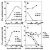
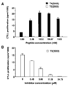
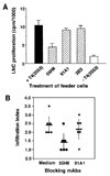
Thyroid hormone-binding (THB) Abs are frequently detected in autoimmune thyroid disorders but it is unknown whether they can exert immunoregulatory effects. We report that a THB mAb recognizing the 5' iodine atom of the outer phenolic ring of thyroxine (T4) can block T cell recognition of the pathogenic thyroglobulin (Tg) peptide (2549-2560) that contains T4 at aa position 2553 (T4(2553)). Following peptide binding to the MHC groove, the THB mAb inhibited activation of the A(k)-restricted, T4(2553)-specific, mouse T cell hybridoma clone 3.47, which does not recognize other T4-containing epitopes or noniodinated peptide analogues. Addition of the same THB mAb to T4(2553)-pulsed splenocytes largely inhibited specific activation of T4(2553)-primed lymph node cells and significantly reduced their capacity to adoptively transfer thyroiditis to naive CBA/J mice. These data demonstrate that some THB Abs can block recognition of iodine-containing Tg epitopes by autoaggressive T cells and support the view that such Abs may influence the development or maintenance of thyroid disease.
Figures


Similar articles
-
Delineation of five thyroglobulin T cell epitopes with pathogenic potential in experimental autoimmune thyroiditis.J Immunol. 2002 Nov 1;169(9):5332-7. doi: 10.4049/jimmunol.169.9.5332. J Immunol. 2002. PMID: 12391254
-
A thyroxine-containing thyroglobulin peptide induces both lymphocytic and granulomatous forms of experimental autoimmune thyroiditis.J Autoimmun. 1997 Dec;10(6):531-40. doi: 10.1006/jaut.1997.0160. J Autoimmun. 1997. PMID: 9451592
-
Enhancing or suppressive effects of antibodies on processing of a pathogenic T cell epitope in thyroglobulin.J Immunol. 1999 Jun 15;162(12):6987-92. J Immunol. 1999. PMID: 10358139
-
T cell mapping of one epitope from thyroglobulin inducing experimental autoimmune thyroiditis (EAT).Int Rev Immunol. 1992;9(2):125-33. doi: 10.3109/08830189209061787. Int Rev Immunol. 1992. PMID: 1283174 Review.
-
On the initial trigger of myasthenia gravis and suppression of the disease by antibodies against the MHC peptide region involved in the presentation of a pathogenic T-cell epitope.Crit Rev Immunol. 2001;21(1-3):1-27. Crit Rev Immunol. 2001. PMID: 11642597 Review.
Cited by
-
B cells limit epitope spreading and reduce severity of EAE induced with PLP peptide in BALB/c mice.J Autoimmun. 2008 Sep;31(2):149-55. doi: 10.1016/j.jaut.2008.04.025. Epub 2008 Jun 9. J Autoimmun. 2008. PMID: 18539432 Free PMC article.
References
-
- Benvenga S, Trimarchi F, Robbins J. Circulating thyroid hormone autoantibodies. J. Endocrinol. Invest. 1987;10:605. - PubMed
-
- Sakata S. Autoimmunity against thyroid hormones. Crit. Rev. Immunol. 1994;14:157. - PubMed
-
- Despres N, Grant AM. Antibody interference in thyroid assays: a potential for clinical misinformation. Clin. Chem. 1998;44:440. - PubMed
-
- Sapin R, Gasser F, Boehn A, Rondeau M. Spuriously high concentration of serum free thyroxine due to anti-triiodothyronine antibodies. Clin. Chem. 1995;41:117. - PubMed
-
- Shimon I, Pariente C, Shlomo-David J, Grossman Z, Sack J. Transient elevation of triiodothyronine caused by triiodothyronine autoantibody associated with acute Epstein-Barr-virus infection. Thyroid. 2003;13:211. - PubMed
Publication types
MeSH terms
Substances
Grants and funding
LinkOut - more resources
Full Text Sources
Molecular Biology Databases
Research Materials
Miscellaneous

