. 2005 Apr;132(7):1499-509.
doi: 10.1242/dev.01719. Epub 2005 Feb 23.

Quail-duck chimeras reveal spatiotemporal plasticity in molecular and histogenic programs of cranial feather development

Affiliations

Quail-duck chimeras reveal spatiotemporal plasticity in molecular and histogenic programs of cranial feather development

B Frank Eames et al. Development. 2005 Apr.

Abstract

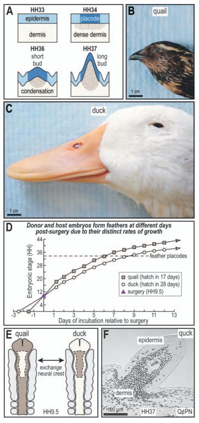
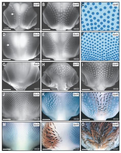
The avian feather complex represents a vivid example of how a developmental module composed of highly integrated molecular and histogenic programs can become rapidly elaborated during the course of evolution. Mechanisms that facilitate this evolutionary diversification may involve the maintenance of plasticity in developmental processes that underlie feather morphogenesis. Feathers arise as discrete buds of mesenchyme and epithelium, which are two embryonic tissues that respectively form dermis and epidermis of the integument. Epithelial-mesenchymal signaling interactions generate feather buds that are neatly arrayed in space and time. The dermis provides spatiotemporal patterning information to the epidermis but precise cellular and molecular mechanisms for generating species-specific differences in feather pattern remain obscure. In the present study, we exploit the quail-duck chimeric system to test the extent to which the dermis regulates the expression of genes required for feather development. Quail and duck have distinct feather patterns and divergent growth rates, and we exchange pre-migratory neural crest cells destined to form the craniofacial dermis between them. We find that donor dermis induces host epidermis to form feather buds according to the spatial pattern and timetable of the donor species by altering the expression of members and targets of the Bone Morphogenetic Protein, Sonic Hedgehog and Delta/Notch pathways. Overall, we demonstrate that there is a great deal of spatiotemporal plasticity inherent in the molecular and histogenic programs of feather development, a property that may have played a generative and regulatory role throughout the evolution of birds.


Figures

Fig. 1
Fig. 1
Quail-duck chimeric system to study cranial feather morphogenesis. (A) Cranial feather buds arise via interactions between the neural crest-derived dermis and the overlying epidermis. At HH33, there is little histological evidence for cranial feather development, but by HH34, epithelial placodes form in the epidermis and the mesenchyme aggregates into dense dermis. By HH36, the feather buds contain a discrete dermal condensation and they begin to rise above the level of the integument. Long buds are present after HH37. (B) Japanese quail and (C) white Pekin duck display considerable differences in the pattern, pigmentation and morphology of their head feathers. (D) Owing to their distinct maturation rates, quail and duck embryos that are stage-matched for surgery subsequently deviate in stage, which provides a potent experimental system with which to identify molecular signals that regulate feather morphogenesis. (E) Neural crest cells were cut either bilaterally (as shown) or unilaterally from the rostral neural tube and exchanged between quail and duck embryos stage-matched at HH9.5. Among other derivatives, these cells are destined to form much of the craniofacial dermis. (F) Chimeric ‘quck’ feather follicles contain duck host epidermis and quail-derived donor dermis stained black with an anti-quail antibody (Q¢PN). Individual quail-derived melanocytes associated with the duck host epidermis are present. Scale bar: 1 cm in B,C; 100 μm in F.
Fig. 2
Fig. 2
The role of neural crest in the spatiotemporal patterning of cranial feather buds. (A) Starting at HH34, quail feather placodes can be seen in one medial and two lateral rows along cranial epidermis (arrow). (B) By HH35, additional rows appear over the eyes. (C) Quail feather buds are relatively large and widely spaced, shown schematically. (D) Duck feather placodes form at HH34 in multiple rows over the eyes, lateral to the midline (arrow). (E) Additional rows appear by HH35. (F) Compared with those of quail, duck feather buds are smaller and spaced closer together, as shown schematically. (G) At HH33, there are no cranial feather buds visible in either duck or quail (not shown). (H) However, HH33 chimeric ‘quck’ prematurely form feather placodes in duck host epidermis. The extent of differentiation, size and spacing of these quck feather buds are more like that observed in I. HH36 quail instead of duck, which is the host species. (J-L) When quck are at HH36, their feathers are like those found on HH39 quail. (M-O) Differences between host and donor feathers are more apparent by HH38, when quck feathers are like those of a quail at HH41. There is unilateral distribution of quail-derived pigment on the duck host, which is coincident with the type of neural crest transplant performed at HH9.5. Scale bar: 2 mm.
Fig. 3
Fig. 3
Cranial neural crest regulates histogenic and molecular programs of feather morphogenesis. (A) At HH33, there are no cranial feather buds in either duck or quail (not shown) as stained histologically with trichrome (TC). (B,C) However, in chimeric ‘quck’ at HH33, the dermis is derived from quail donor neural crest (Q¢PN positive, black cells), which is on a faster timetable for development and induces premature formation of feather buds in duck host epidermis. The extent of differentiation, size and spacing of these quck feather buds are more like that observed in D, a HH36 control quail instead of an HH33 duck, which is the host species. (E) Control duck do not form short feather buds until HH36. (F) In situ hybridization analyses reveal that molecular markers of feather development such as bmp4 are not expressed in the capital tracts of control quail and duck prior to HH34. (G,H) However, chimeric quck at HH33 express bmp4 in cranial feather mesenchyme, which is equivalent to that observed for control quail at HH36. (I,J) HH33 quck express bmp2 in the epithelium and mesenchyme like HH36 quail. (K,L) HH33 quck express follistatin in the epithelium and mesenchyme like HH36 quail. (M,N) HH33 quck express bmpr1a in the epithelium and mesenchyme like HH36 quail. (O,P) HH33 quck express shh in the epithelium like HH36 quail. (Q,R) HH33 quck express ptc in the epithelium and mesenchyme like HH36 quail. (S,T) HH33 quck express delta1 in the mesenchyme like HH36 quail. (U,V) HH33 quck express notch1 in the epithelium and mesenchyme like HH36 quail. (W) At HH31, there are no epidermal placodes or underlying dense dermis present in control duck or quail (not shown). (X) These do not appear in control quail and duck (not shown) until HH34 (arrow). (Y,Z) However, in chimeric quck at HH31, quail donor neural crest-derived mesenchyme has given rise to dense dermis (Q¢PN positive) and has induced epidermal placodes (arrows) of duck host origin (Q¢PN negative). (A′,B′) In situ hybridization analyses reveal that bmp4 and bmp2 are expressed in quail donor-derived mesenchyme prematurely at HH31, whereas normally they would not be expressed until HH34 (arrows). Other mesenchymal and epithelial molecular markers of feather morphogenesis are not yet detected except for bmpr1a and notch1, which are expressed continuously from at least HH29 in most tissues (data not shown). Scale bar: 100 μm.
Fig. 4
Fig. 4
Donor neural crest can also delay molecular and histogenic programs of cranial feather development. (A) Duck cranial neural crest cells follow their own timetable for differentiation when transplanted into quail hosts. Resultant duail chimeras collected at HH36 contain feather buds as well as epidermal regions that lack placodes (broken outline). The absence of epidermal placodes is equivalent to that observed on control embryos at HH33 (compare with Fig. 2G). (B,C) The presence and absence of cranial feather buds can be seen in sections stained histologically with trichrome (TC). (D) Immunohistochemical analyses using an anti-quail antibody confirm that wherever placodes are present, the dermis is derived from quail host neural crest (Q¢PN positive, black cells). (E) By contrast, regions that lack placodes contain dermis derived from the duck donor (Q¢PN negative). (F,G) Bmp4 is expressed in mesenchyme derived from the quail host but is not yet detected in mesenchyme of duck donor origin. (H,I) Shh is expressed in the epithelium overlying dermis from the quail host but not over dermis derived from the duck donor. (J,K) Delta1 is detected in quail host dermis but not in duck donor-derived dermis. (L) Duail chimeras collected at HH39 have normal long feather buds alongside areas containing short feather buds like those observed on HH36 controls. (M) Immunohistochemical analyses confirm that the short feather buds, which are like those of a HH36 duck instead of a HH39 quail, are derived from duck donor neural crest (Q¢PN-negative). (N) Long feather buds composed of quail host epidermis (Q¢PN-positive) and duck donor dermis (Q¢PN-negative; arrow) are present in duail chimeras at HH42. Scale bar: 1 mm in A; 100 μm in B-K; 1 mm in L; 50 μm in M; 100 μm in N.
Fig. 5
Fig. 5
Quail-duck chimeras reveal plasticity in cranial feather development. Bars represent stages when histogenic and molecular events are initiated in controls versus chimeras.

References

    1. Alberch P. Problems with the interpretation of developmental sequences. System. Zool. 1985;34:46–58.
    1. Albrecht UEG, Helms JA, Lin H. Visualization of gene expression patterns by in situ hybridization. In: Daston GP, editor. Molecular and Cellular Methods in Developmental Toxicology. CRC Press; Boca Raton, FL: 1997. pp. 23–48.
    1. Ashique AM, Fu K, Richman JM. Signalling via type IA and type IB bone morphogenetic protein receptors (BMPR) regulates intramembranous bone formation, chondrogenesis and feather formation in the chicken embryo. Int. J. Dev. Biol. 2002;46:243–253. - PubMed
    1. Atit R, Conlon RA, Niswander L. EGF signaling patterns the feather array by promoting the interbud fate. Dev. Cell. 2003;4:231–240. - PubMed
    1. Bee J, Thorogood P. The role of tissue interactions in the skeletogenic differentiation of avian neural crest cells. Dev. Biol. 1980;78:47–66. - PubMed

Publication types

MeSH terms