. 2005 May;16(5):2325-38.
doi: 10.1091/mbc.e04-11-0996. Epub 2005 Feb 23.

Dissociation of the Nuf2-Ndc80 complex releases centromeres from the spindle-pole body during meiotic prophase in fission yeast

Affiliations

Dissociation of the Nuf2-Ndc80 complex releases centromeres from the spindle-pole body during meiotic prophase in fission yeast

Haruhiko Asakawa et al. Mol Biol Cell. 2005 May.

Abstract

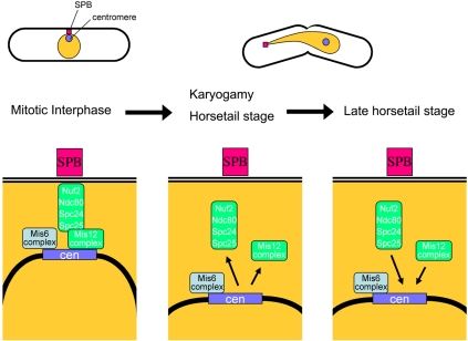
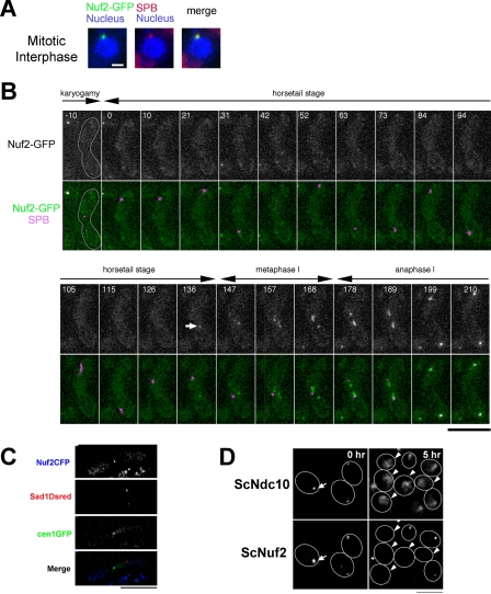
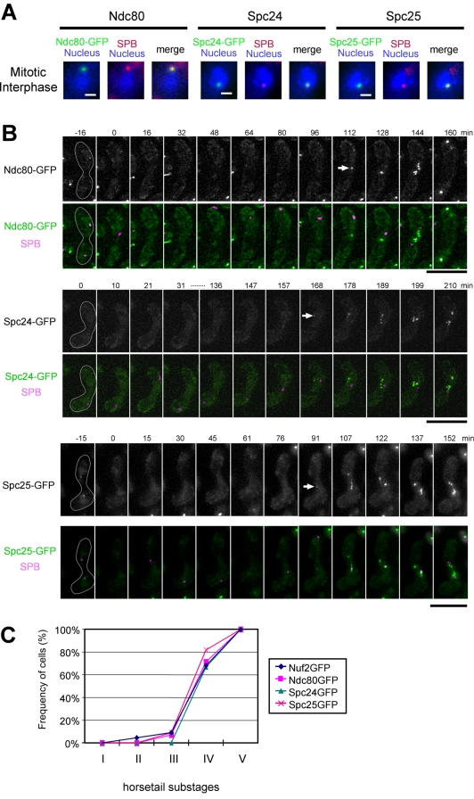
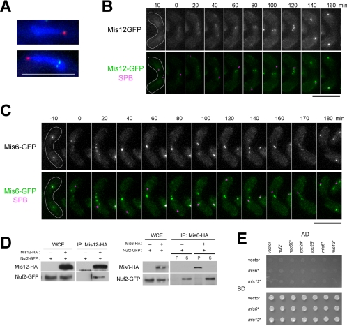
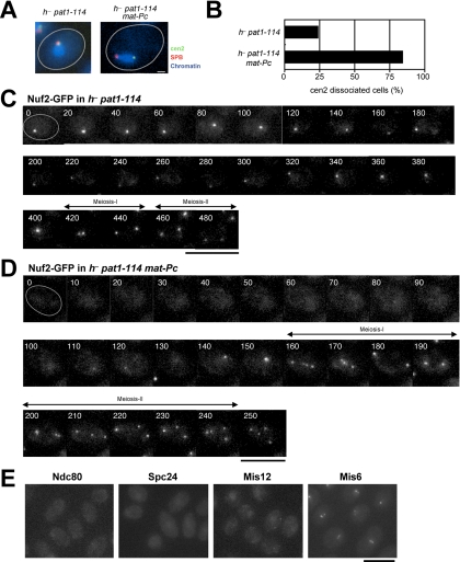
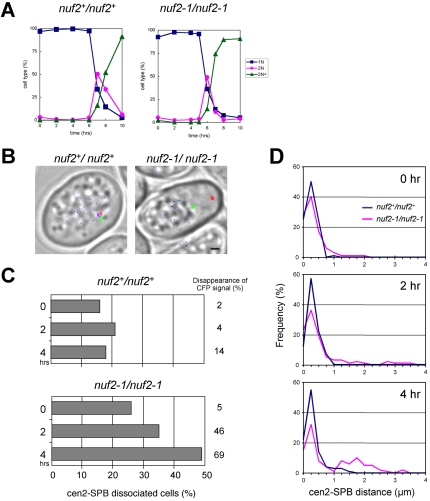
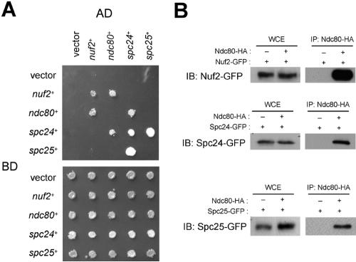
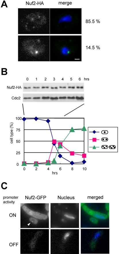
In the fission yeast Schizosaccharomyces pombe, centromeres remain clustered at the spindle-pole body (SPB) during mitotic interphase. In contrast, during meiotic prophase centromeres dissociate from the SPB. Here we examined the behavior of centromere proteins in living meiotic cells of S. pombe. We show that the Nuf2-Ndc80 complex proteins (Nuf2, Ndc80, Spc24, and Spc25) disappear from the centromere in meiotic prophase when the centromeres are separated from the SPB. The centromere protein Mis12 also dissociates during meiotic prophase; however, Mis6 remains throughout meiosis. When cells are induced to meiosis by inactivation of Pat1 kinase (a key negative regulator of meiosis), centromeres remain associated with the SPB during meiotic prophase. However, inactivation of Nuf2 by a mutation causes the release of centromeres from the SPB in pat1 mutant cells, suggesting that the Nuf2-Ndc80 complex connects centromeres to the SPB. We further found that removal of the Nuf2-Ndc80 complex from the centromere and centromere-SPB dissociation are caused by mating pheromone signaling. Because pat1 mutant cells also show aberrant chromosome segregation in the first meiotic division and this aberration is compensated by mating pheromone signaling, dissociation of the Nuf2-Ndc80 complex may be associated with remodeling of the kinetochore for meiotic chromosome segregation.


Figures

Figure 8.
Figure 8.
The S. pombe kinetochore structure. In mitotic interphase, centromeres locate near the SPB along with kinetochore complexes, including Nuf2-Ndc80 complex, Mis12 complex and Mis6 complex. During karyogamy and the horsetail stage, Nuf2-Ndc80 complex and Mis12 disappear from the centromere leading to the centromere-SPB dissociation. At the late horsetail stage, Mis12 complex and Nuf2-Ndc80 complex reappear at the centromere.
Figure 1.
Figure 1.
Behavior of Nuf2 during meiosis. (A) Nuf2-GFP (green) and Sad1-DsRed (red) in mitotically growing cells of S. pombe strain (HA252–4A). The nucleus was stained with Hoechst33342 (blue). Bar, 1 μm. (B) Live cell observation of S. pombe Nuf2-GFP during karyogamy and meiotic prophase. HA252–4A strain was incubated on sporulation medium, and time-lapse images of living zygote cell were acquired. Nuf2-GFP (green) and Sad1-DsRed (magenta) are shown. Time 0 indicates when the Sad1-DsRed spots are fused. Although Nuf2-GFP disappears from centromeres, weak GFP signals are sometime observed in cytoplasm (e.g., frames at 31 and 42 min); such cytoplasmic signals can be distinguished from centromeric signals because they do not follow horsetail nuclear movements. Bar, 10 μm. (C) Localization of Nuf2 at the centromere. The HA525 strain was incubated on sporulation medium. Nuf2-CFP (blue), cen1-GFP (green), and Sad1-DsRed (red). Bar, 10 μm. (D) Observation of the S. cerevisiae Nuf2 protein during meiotic prophase. S. cerevisiae NKY278 strain was taken at 0 and 5 h after induction of meiosis and then prepared for immunostaining. Ndc10 protein was stained as a centromere marker. Bar, 15 μm.
Figure 3.
Figure 3.
Behaviors of Nuf2-Ndc80 complex proteins during karyogamy and meiotic prophase. (A) Observation of the S. pombe Ndc80, Spc24, and Spc25 proteins. Nuf2-interactors (green) and Sad1-DsRed (red) were observed in mitotically growing cells (HA205–2A, HA229–11C, and HA485–1D strains). The nucleus was stained with Hoechst33342 (blue). Bar, 1 μm. (B) Live cell observation of Ndc80, Spc24, and Spc25 during karyogamy and meiotic prophase. HA205–2A, HA229–11A, or HA485–1D strain was incubated on sporulation medium, and time-lapse observations of living zygote cells were performed as described in Figure 1. Ndc80-GFP, Spc24-GFP, or Spc25-GFP (green), and Sad1-DsRed (magenta) are shown in merged images. Bar, 10 μm. (C) Kinetics of the behavior of Nuf2-Ndc80 complex proteins. Time-lapse images of cells expressing Nuf2-GFP, Ndc80-GFP, Spc24-GFP, or Spc25-GFP were recorded at 5-min intervals. The horsetail stage was divided equally into five substages. In each substage, the number of time points when a spot signal of GFP was observed was counted and divided by the total number of time points to calculate the frequency of positive GFP signals. The percentages were plotted as a time course of the substages. Numbers of living cells examined: 22 for Nuf2-GFP, 14 for Ndc80-GFP, 12 for Spc24-GFP, and 11 for Spc25-GFP.
Figure 4.
Figure 4.
Behavior of Mis12 and Mis6. (A) Localization of Mis12 or Mis6 in meiotic prophase. The Mis12-GFP (HA259–1D, top) and Mis6-GFP strains (HA260–7D, bottom) were incubated on sporulation media, and zygotic cells were observed. Typical horsetail nuclei are shown. The Mis12-GFP and Mis6-GFP were shown in green. The SPB was visualized by Sad1-DsRed (red), and the nuclei were stained with Hoechst33342 (blue). (B and C) Behavior of Mis12 and Mis6 during karyogamy and meiotic prophase. Living zygotes of the Mis12-GFP (HA259–1D) or Mis6-GFP strains (HA260–7D) were observed every 10 min in sporulation medium. Mis12-GFP or Mis6-GFP (green) and Sad1-DsRed (magenta) are also shown in merged images. Bars, 10 μm. (D) Immunoprecipitation assay between Mis12 and Nuf2 (left) and between Mis6 and Nuf2 (right). Extracts of Mis12 and Mis12-HA cells expressing Nuf2-GFP (HA282–3D and HA252–12B) and Mis6 and Mis6-HA cells expressing Nuf2-GFP (HA380-11C and HA252–12B) were incubated with anti-HA beads, and precipitates (IP) were subjected to immunoblot analysis using anti-GFP antibody. Whole-cell extracts were also analyzed (WCE). The extracts were prepared from growing cultures. In the right panel, P, precipitate; S, supernatant. (E) Yeast two-hybrid assay between Mis6 or Mis12 and Nuf2-Ndc80 complex proteins. Yeast strains were transformed with the combinations of plasmids as indicated, spotted onto selective (top) and nonselective media (bottom), and incubated for 3 d.
Figure 5.
Figure 5.
Behavior of Nuf2 in pat1 mutant cells upon mating pheromone signaling. (A) Location of centromeres and SPB in living pat1 mutant cells in the absence or presence of the c-type mat gene. The strains HA499-1C (h pat1-114) or HA495–9B (h pat1-114 plus mat-Pc) were arrested in G1 by nitrogen starvation at 26°C. In these strains the centromere region of chromosome II was visualized using GFP-LacI (which recognizes the lacO sequence) and the SPB was visualized using Sad1-DsRed. The nuclei were stained with Hoechst33342 (blue). Bar, 10 μm. (B) Statistical analysis of the detachment of a centromere (cen2) from the SPB in A. n = 100 in each strain. (C) Behavior of Nuf2 in pat1-induced meiosis. h pat1-114 cells expressing Nuf2-GFP (HA309–11C) were arrested in G1 by nitrogen starvation at a permissive temperature of 26°C, and the Nuf2-GFP was then observed after transfer to 34°C. Numbers in each frame indicate the time in minutes after the temperature shift. Bar, 10 μm. (D) Behavior of Nuf2 in pat1 mutant cells in the presence of the c-type mat gene. A strain HAR3021-1 (h pat1-114 nuf2+-GFP strain harboring mat-Pc) was observed as described in C. Bar, 10 μm. (E) Behavior of Ndc80, Spc24, Mis12, and Mis6 in pat1 mutant cells in the presence of the c-type mat gene. Ndc80-GFP, Spc24-GFP, Mis12-GFP, and Mis6-GFP were observed in h pat1-114 strains harboring the mat-Pc gene (HAR3041-1, HAR3031-1, HA3505C-1, and CRLa59-1) after the induction of G1 arrest by nitrogen starvation at 26°C. Bar, 10 μm.
Figure 7.
Figure 7.
Dissociation of centromeres by nuf2-1 mutation in pat1-induced meiotic prophase. (A) Progression of meiosis in nuf2+/nuf2+ and nuf2-1/nuf2-1 strains in the background of pat1ts mutation (HA529 and HA530, respectively). Both strains were arrested in G1 by nitrogen starvation at 26°C and then transferred to 34°C. Nuclear division was monitored by staining the nuclei with DAPI. (B) Localization of centromeres, SPB and Nuf2. Both strains were observed 4 h after the temperature shift in A. The peri-centromeric region of chromosome II (cen2) visualized by lacO/GFP-LacI method (Yamamoto and Hiraoka, 2003; green), SPB visualized by Sad1-DsRed (red), and Nuf2-CFP (blue) are shown as merged images with bright-field micrographs. Bar, 1 μm. (C) Frequency of the centromere-SPB separation. The distance between the centromere (cen2) and SPB was measured in cells of each strain at indicated time points after the temperature shift (0 h) in A. At least 100 cells were observed at each time point. The cell in which the cen2-SPB distance was >0.55 μm is shown as a cen2-SPB dissociated cell. Distances between cen2 and SPB for two strains were further compared by the Mann-Whitney U test. p values, 0.79 at 0 h, 0.69 at 2 h, and <0.0001 at 4 h. The number at the right of each graph indicates the percentage of cells with no detectable CFP signals of Nuf2 or Nuf2–1 protein. (D) Distribution of the centromere-SPB distance. Histogram of the cen2-SPB distance measured in C was made with 0.25-μm intervals and plotted for nuf2+ (blue) and nuf2-1 (red) at 0, 2, and 4 h.
Figure 2.
Figure 2.
Interactions of Nuf2-Ndc80 complex proteins. (A) Yeast two-hybrid assay of the Nuf2-Ndc80 complex. Yeast cells were transformed with the combinations of plasmids as indicated, spotted onto selective (top) and nonselective media (bottom), and incubated for 3 d. SD/-Leu, -Trp, -His, and -Ade medium, containing 1 mM 3-amino-1,2,4-triazole (3-AT) was used as the selective medium. (B) Immunoprecipitation assay of the Nuf2-Ndc80 complex. Extracts expressing Nuf2-GFP (HA252–12B and HA374–2C, top panel), Spc24-GFP (HA229–1C and HA329–3A, middle panel), or Spc25-GFP (HA484–1A and HA484–2C, bottom panel) were prepared in the absence or presence of Ndc80-HA (Ndc80-HA “−” or “+”, respectively). All extracts were prepared from growing cultures and incubated with anti-HA beads. The precipitates (IP) and whole cell extracts (WCE) were analyzed by Western blotting using anti-GFP antibody.
Figure 6.
Figure 6.
(A) Immunofluorescence staining of Nuf2-HA protein in h pat1-114 with mat-Pc (strain HA626-4D). Cells were fixed after the induction of G1 arrest by nitrogen starvation and subjected to immunofluorescence. More than 200 cells were observed to detect Nuf2-HA. Bar, 1 μm. (B) Expression of Nuf2 during meiosis. Cells of strain HA626–4D were arrested in G1 by nitrogen starvation and were induced to undergo synchronous meiosis by shifting the temperature as described in the legend to Figure 5. Cell extracts were prepared and subjected to immunoblot analysis with anti-HA antibody. As a loading control, Cdc2 was detected by anti-Cdc2 antibody. The kinetics of nuclear division were also monitored by staining nuclei with DAPI. (C) Localization of ectopically expressed Nuf2. Cells of strain HA478–1C, which express Nuf2-GFP under the thiamine repressible nmt1 promoter, were incubated on sporulation medium with (lower) or without thiamine (upper), and meiotic prophase cells were observed. Nuclei were stained with Hoechst-33342. The arrowhead indicates a mitotic interphase cell.

References

    1. Appelgren, H., Kniola, B., and Ekwall, K. (2003). Distinct centromere domain structures with separate functions demonstrated in live fission yeast cells. J. Cell Sci. 116, 4035–4042. - PubMed
    1. Beach, D., Rodgers, L., and Gould, J. (1985). RAN1+ controls the transition from mitotic division to meiosis in fission yeast. Curr. Genet. 10, 297–311. - PubMed
    1. Bernard, P., Maure, J. F., and Javerzat, J. P. (2001). Fission yeast Bub1 is essential in setting up the meiotic pattern of chromosome segregation. Nat. Cell Biol. 3, 522–526. - PubMed
    1. Bharadwaj, R., Qi, W., and Yu, H. (2004). Identification of two novel components of the human NDC80 kinetochore complex. J. Biol. Chem. 279, 13076–13085. - PubMed
    1. Chikashige, Y., Ding, D. Q., Funabiki, H., Haraguchi, T., Mashiko, S., Yanagida, M., and Hiraoka, Y. (1994). Telomere-led premeiotic chromosome movement in fission yeast. Science 264, 270–273. - PubMed

Publication types

MeSH terms

LinkOut - more resources