. 2005 May;16(5):2248-62.
doi: 10.1091/mbc.e04-07-0639. Epub 2005 Feb 23.

Identification and characterization of a novel alpha-kinase with a von Willebrand factor A-like motif localized to the contractile vacuole and Golgi complex in Dictyostelium discoideum

Affiliations

Identification and characterization of a novel alpha-kinase with a von Willebrand factor A-like motif localized to the contractile vacuole and Golgi complex in Dictyostelium discoideum

Venkaiah Betapudi et al. Mol Biol Cell. 2005 May.

Abstract

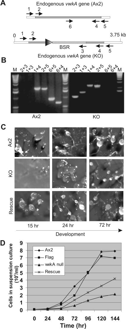
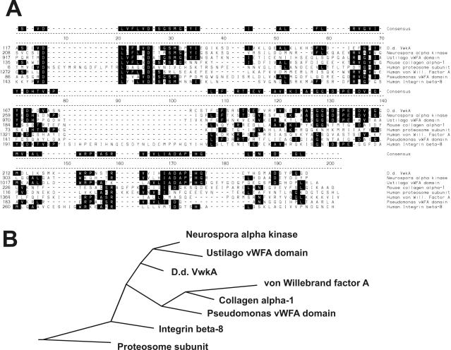
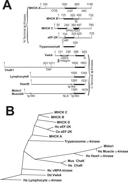
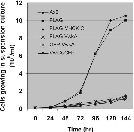
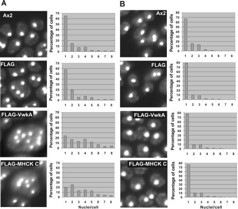
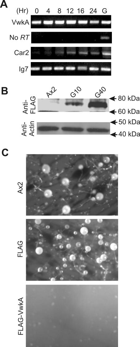
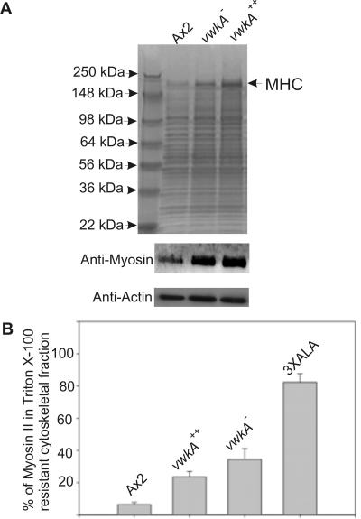
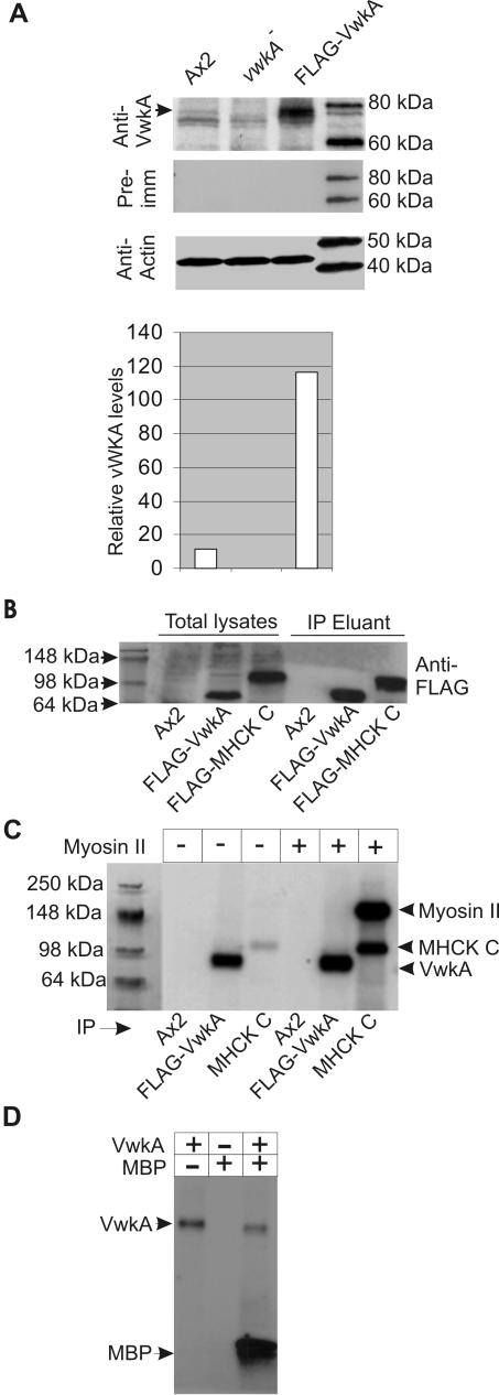
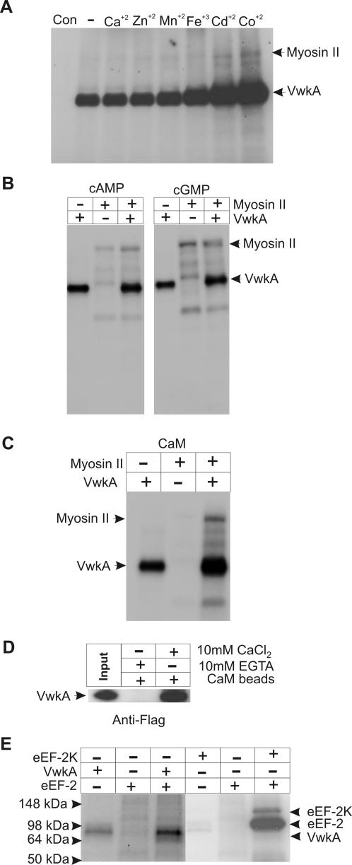
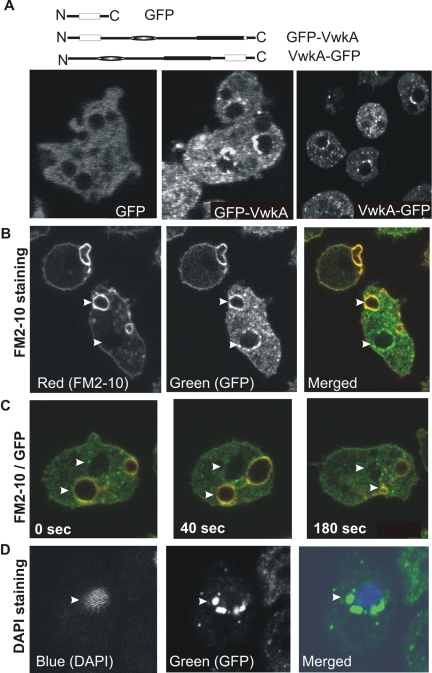
We have identified a new protein kinase in Dictyostelium discoideum that carries the same conserved class of "alpha-kinase" catalytic domain as reported previously in myosin heavy chain kinases (MHCKs) in this amoeba but that has a completely novel domain organization. The protein contains an N-terminal von Willebrand factor A (vWFA)-like motif and is therefore named VwkA. Manipulation of VwkA expression level via high copy number plasmids (VwkA++ cells) or gene disruption (vwkA null cells) results in an array of cellular defects, including impaired growth and multinucleation in suspension culture, impaired development, and alterations in myosin II abundance and assembly. Despite sequence similarity to MHCKs, the purified protein failed to phosphorylate myosin II in vitro. Autophosphorylation activity, however, was enhanced by calcium/calmodulin, and the enzyme can be precipitated from cellular lysates with calmodulin-agarose, suggesting that VwkA may directly bind calmodulin. VwkA is cytosolic in distribution but enriched on the membranes of the contractile vacuole and Golgi-like structures in the cell. We propose that VwkA likely acts indirectly to influence myosin II abundance and assembly behavior and possibly has broader roles than previously characterized alpha kinases in this organism, which all seem to be MHCKs.


Figures

Figure 6.
Figure 6.
Generation of vwkA null cells and evaluation of development. (A) Strategy for the deletion of vwkA gene. A gene-targeting cassette carrying a blasticidin selection marker (BSR; lower schematic image) was transfected into Ax2 cells, and clonal blasticidin-resistant cell lines were isolated. PCR analysis was performed with genomic DNA to identify candidate vwkA null lines, followed by extensive PCR analysis with indicated primers 1–6 as indicated by arrows. Sequences of these primers are given in Materials and Methods. PCR reactions were configured to discriminate between the wild-type locus in parental Ax2 cells (top schematic image) and the expected locus organization resulting from double crossover gene targeting to create a vwkA null (lower schematic). (B) Example of genomic PCR analysis of one vwkA null cell line. PCR amplifications performed as described in previously (Betapudi et al., 2004) were subjected to electrophoresis on 1.2% agarose gel. M indicates 1-kb DNA ladder marker. Ax2 lanes indicate PCR results obtained with parental Ax2 genomic DNA, KO indicates PCR results obtained with genomic DNA from a vwkA null line. PCR results confirm the lack of a complete vwkA gene in the vwkA null lines. For example, the vwkA null line genomic DNA fails to yield a product with primers 6 + 5 or 6 + 4, and with primers 1 + 4 or 2 + 5, the vwkA null displays a large product than Ax2, due to the replacement of the central part of the vwkA gene with the BSR cassette. (C) Developmental defects of vwkA null cells and rescued lines. The parental Ax2 cells, vwkA null cells, and vwkA null cells transfected with a FLAG-tagged VwkA expression construct (selected with G418 at a low concentration of 5 μg/ml) were grown in plastic petri plates and then collected and developed as in Figure 5. Images of the developing cells were taken at the indicated time points during development. (D) Suspension growth defects of vwkA null cells and rescued line. The parental Ax2 cells, vwkA null cells, and vwkA null cells transfected with a FLAG-tagged VwkA expression construct (selected with G418 at 5 μg/ml) were grown in plastic petri plates and then transferred to flasks containing HL5 for suspension growth. Cell densities were determined daily via hemocytometer counts. The “Flag” sample refers to growth of Ax2 cells transfected with the empty pTX-FLAG vector and grown in 5 μg/ml G418. The “rescue” sample refers to vwkA null cells transfected with the FLAG-VwkA construct and grown in 5 μg/ml G418.
Figure 1.
Figure 1.
The vWFA/β-integrin motif of VwkA. (A) ClustalW alignment of the vWFA domain of VwkA with other vWFA domain family members. Consensus residues are defined as residues identical in at least three of the sequences and are shaded. (B) Phylogenetic tree based upon ClustalW alignment. Alignment and tree produced with the DNAStar program MegAlign. GenBank accession numbers for included sequences are as follows: Neurospora α-kinase (discussed later in paper), XP_323573; Ustilago vWFA domain containing protein, EAK82057; mouse collagen α-1, Q60847; human proteosome 26S subunit Rpn10, P55036; human von Willebrand factor A (motif A1), P04275; Pseudomonas vWFA domain protein, NP-744179; and human β-integrin 8, P26012.
Figure 2.
Figure 2.
(A) Schematic representation of α-kinase family member domain organization. The N-terminal and C-terminal kinase subfamilies are indicated by brackets. WD refers to the WD repeat domain (gray box), and coil refers to segments with strongly predicted coiled-coil character (cross-filled box). N refers to polyasparagine-rich sequence; SPNQ refers to serine, asparagines, proline, and glutamic acid-rich sequence; and S refers to a serine-rich sequence. A possible PEST domain in MHCK C (proline, glutamic acid, serine, and threonine-rich) is indicated, and CaM refers to Ca+2-calmodulin binding motif. Distorted circle in VwkA indicates vWFA/β-integrin motif. NLS refers to nuclear localization signal, Ig-like refers to immunoglobulin-like, and TRP indicates transient receptor potential ion channel domain. The amino acid numbers shown for each correspond to the following accession numbers: MHCK A (A55532), MHCK B (AAB50136), MHCK C (AAC31918), human eEF2K (NP_037434), mouse Chak1 (AFF73131), human LymphocyteK (AAK94675) human HeartK (NP_443179), mouse Midori/MuscleK (NP_473426), and Trypanosome kinase (AC079815). (B) Phylogenetic tree of α-kinase catalytic domains. ClustalW alignments and phylogenetic tree were performed with the DNAStar software package. Protein sequence sources listed in A, but alignments also included the D. discoideum genomic sequence for MHCK D (our unpublished data and from genome project), and an N. crassa sequence for an α-kinase (Nc vWFA kinase; XP_328675) related in domain organization to Dictyostelium VwkA.
Figure 3.
Figure 3.
Cell growth kinetics of VwkA-overexpressing cell lines in suspension culture. Cells were collected from petri dish culture and transferred to flasks rotating at 200 rpm at 22°C in HL5 medium at an initial density of 1 × 10⁵ cells/ml. Cell lines evaluated were parental Ax2, cells expressing empty FLAG-tag vector (FLAG), FLAG-tagged VwkA (FLAG-VwkA), FLAG-tagged MHCK C (FLAG-MHCK-C), N-terminal GFP-tagged VwkA (GFP-VwkA), and C-terminal tagged VwkA (VwkA-GFP). All of the stable cells expressing fusion proteins or FLAG-vector were grown in the HL5 with G418 at 10 μg/ml. Cell density was determined by hemocytometer counts every 24 h.
Figure 4.
Figure 4.
Nuclear staining of cells. (A) DAPI staining of cells growing in suspension culture. Cells growing in suspension culture were transferred to glass-bottomed microscope chambers, allowed to attach for 10 min, and then fixed and DAPI stained as described in Materials and Methods. (B) DAPI staining of cells growing in plastic petri plates. Cells growing in plastic petri plates to near confluent were collected, fixed, and stained as described in Materials and Methods. Cells were scored with respect to nuclei/cell as presented. Each graph represents score of 300 cells from several fields of view.
Figure 5.
Figure 5.
Development of VwkA-overexpressing cells. (A) RT-PCR analysis of vwkA gene expression during development of Ax2 cells. Ax2 cells growing in suspension culture were collected by centrifugation followed by washing with starvation buffer. The cell pellet was resuspended in starvation buffer at 2 × 10⁸ cells/ml and layered on presoaked Whatman filter paper in starvation buffer placed on top of agarose plates made in starvation buffer. RNA was isolated at indicated times. RT-PCR was performed with vwkA-specific primers in the presence of reverse transcriptase (top) or as a control, without reverse transcriptase (second panel from top). G represents control PCR performed with genomic DNA as template. PCR mixtures were subjected to electrophoresis on 1% agarose gel. As referenced in Materials and Methods, Car2 is a control developmentally expressed gene, and Ig7 is a control constitutively expressed gene. (B) Analysis of FLAG-VwkA overexpression levels. Total cell lysates were subjected to Western blot analyses by using FLAG antibodies. These cell lysates also were subjected to Western blot analysis by using actin antibodies as a loading control. Left lane, parental Ax2 cells; middle lane, FLAG-VwkA cells growing in medium with G-418 at 10 μg/ml; and right lane, FLAG-VwkA cells growing in medium with G418 at 40 μg/ml. (C) Development of FLAG-VwkA–overexpressing cells. Parental Ax2 and stable transfected cell lines expressing empty FLAG vector or FLAG-VwkA fusion protein were grown in the presence of G418 at 40 μg/ml and then subjected to development as described above. Images were taken after 72 h of development.
Figure 7.
Figure 7.
Effect of VwkA expression level on myosin II expression and assembly. (A) SDS-PAGE analysis of total cell extracts. Total cell extracts were made from VwkA++, vwkA null, and parental Ax2 cells grown in plastic petri plates by passage through Nucleopore membranes. Equal quantities of total protein from each sample were subjected to SDS-PAGE, followed by Coomassie Blue staining. Arrow indicates MHC band, further confirmed by performing Western blot analysis by using anti-myosin II antibody My4 (Flicker et al., 1985; Peltz et al., 1985) as shown in second panel. The myosin II Western blot was stripped and reprobed with anti-actin antisera to confirm equal loading of lysates samples. (B) Myosin II levels in Triton X-100–insoluble cytoskeletal ghosts. Triton X-100 lysates were fractionated via centrifugation, and percentage of MHC present in the cytoskeletal ghost was quantified via SDS-PAGE, Coomassie staining, and densitometry. Error bars represent SEM, and n = 8 independent samples for each bar.
Figure 8.
Figure 8.
Immunopurification of FLAG-VwkA and protein kinase activity. (A) Western blot analysis of FLAG-VwkA overexpression. Total cell lysates were subjected to SDS-PAGE and Western blot analysis with anti-VwkA peptide antisera. Ax2 lysates reveal a weak but reproducible band at ∼75 kDa (arrow), which is absent in vwkA null cell lysates. FLAG-VwkA–overexpressing cells (grown in G418 at 10 μg/ml) display a robust signal at this position. Western blot samples also were probed with anti-actin antibodies as a loading control. Densitometric analysis of Western blots indicates ∼11-fold overexpression of FLAG-VwkA relative to Ax2. (B) Western blot analysis of immunoprecipitates. Western blot analyses were performed with anti-FLAG antibodies and either Ax2 lysates (negative control), FLAG-VwkA lysates, or FLAG-MHCK-C lysates. The right three lanes indicate signal from material that was immunopurified and eluted from beads with competing FLAG peptide. (C) Autoradiogram of in vitro phosphorylation assays with immunopurified FLAG-VwkA using myosin II as a substrate. Reactions were performed in 20-μl volume containing 1 μM myosin II purified from Dictyostelium and incubated at 22°C for 20 min in a mixture containing 2 mM MgCl2 and 0.5 mM [32P]γ-ATP (1 μCi). Reaction mixtures were stopped by addition of SDS-sample buffer and subjected to SDS-PAGE. Dried gels were exposed to x-ray films. (D) VwkA phosphorylates MBP efficiently. In vitro phosphorylation assay was performed as described in C for myosin II except using 5 μM MBP instead of myosin II in the reaction mixture.
Figure 9.
Figure 9.
Evaluation of VwkA kinase substrates and activation. (A) Effect of metal ions on phosphorylation of myosin II by FLAG-VwkA. Phosphorylation assays performed as in previous figure but with addition of 50 μM CaCl2, ZnCl2, MnCl2, Fe2Cl3, CdCl2, or CoCl2, as indicated, with myosin II as a substrate. (B) Effect of cAMP and cGMP on in vitro phosphorylation of myosin II by FLAG-VwkA. Assays performed as in previous figure, with addition of 0.5 mM cAMP or 0.5 mM cGMP, as indicated, with myosin II as substrate. (C) In vitro phosphorylation of myosin II in the presence of CaM. Phosphorylation assays were carried out using the purified FLAG-VwkA protein in the presence of calcium/calmodulin. (D) FLAG-VwkA is precipitated from crude cell lysates by CaM-agarose. Total lysates from FLAG-VwkA cells were incubated with CaM-agarose beads in the presence of either EGTA or CaCl2. Beads were washed twice in the same solution and then boiled in SDS-PAGE sample buffer, and solubilized proteins were subjected to SDS-PAGE and Western blot analysis by using FLAG antibodies. Left lane indicates signal from equivalent amount of input total cell lysate. (E) eEF-2 is not a substrate for VwkA in vitro. In vitro phosphorylation assays were performed with either mammalian eEF-2K or FLAG-VwkA and purified mammalian eEF-2 as substrate as described by Ryazanov et al. (1997). The eEF-2 and eEF-2 kinase samples were generously provided by Dr. Alexy Ryazanov.
Figure 10.
Figure 10.
Analysis of VwkA subcellular localization. (A) Schematic organization of GFP constructs for imaging. Cells expressing empty GFP vector displayed diffuse GFP fluorescence throughout the cytosol, whereas cells expressing the GFP-VwkA fusion (GFP at N terminus) and the VwkA-GFP (GFP at C terminus) both displayed punctate fluorescence throughout the cytosol in confocal sections. Cell lines growing in plastic petri plates were collected by centrifugation at low speed, washed with starvation buffer, and seeded into glass-bottomed microscopic chambers for live cell imaging via confocal microscopy. Images were collected using a 100× objective on a Zeiss LSM510. (B) Staining of contractile vacuoles with FM2-10 dye: GFP-VwkA cells seeded into glass bottom microscopic chambers as described above were stained with FM2-10 dye and images were collected starting 2 min after addition of dye by exciting at 488 nm. Time-lapse series of images were collected in parallel with red and green emission filters to obtain FM2-10 and GFP emission patterns, respectively. See Supplemental Movie files VwkA-GFP-FM-1.mov and VwkA-GFP-FM-2.mov for additional examples. (C) Contractions of organelles localized with GFP-VwkA. Time-lapse series of GFP-VwkA cell stained with FM2-10, dual image of GFP and FM2–10 signal. Series displays association of GFP signal adjacent to a noncontractile structure (top arrowhead; identified as the nucleus in data below) and GFP signal colocalizing with a contractile vacuole (bottom arrowhead). (D) DAPI staining of cells expressing VwkA-GFP: Cells expressing VwkA-GFP were stained with DAPI as described in text. See Supplemental Movie file VwkA-GFP-DAPI.mov for z-section of fixed cells displaying additional examples.

References

    1. Aarts, M., Iihara, K., Wei, W. L., Xiong, Z. G., Arundine, M., Cerwinski, W., MacDonald, J. F., and Tymianski, M. (2003). A key role for TRPM7 channels in anoxic neuronal death. Cell 115, 863–877. - PubMed
    1. Adachi, H., Hasebe, T., Yoshinaga, K., Ohta, T., and Sutoh, K. (1994). Isolation of Dictyostelium discoideum cytokinesis mutants by restriction enzyme-mediated integration of the blasticidin S resistance marker. Biochem. Biophys. Res. Commun. 205, 1808–1814. - PubMed
    1. Becker, M., Matzner, M., and Gerisch, G. (1999). Drainin required for membrane fusion of the contractile vacuole in Dictyostelium is the prototype of a protein family also represented in man. EMBO J. 18, 3305–3316. - PMC - PubMed
    1. Betapudi, V., Shoebotham, K., and Egelhoff, T. T. (2004). Generation of double gene disruptions in Dictyostelium discoideum using a single antibiotic marker selection. Biotechniques 36, 106–112. - PubMed
    1. Buxton, D. B., Golomb, E., and Adelstein, R. S. (2003). Induction of nonmuscle myosin heavy chain II-C by butyrate in RAW 264.7 mouse macrophages. J. Biol. Chem. 278, 15449–15455. - PubMed

Publication types

MeSH terms