Distinct roles for lateral and medial anterior prefrontal cortex in contextual recollection
- PMID: 15728761
- PMCID: PMC3838933
- DOI: 10.1152/jn.01200.2004
Distinct roles for lateral and medial anterior prefrontal cortex in contextual recollection
Abstract
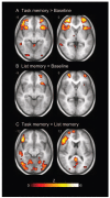
A key feature of human recollection is the ability to remember details of the context in which events were experienced, as well as details of the events themselves. Previous studies have implicated a number of regions of prefrontal cortex in contextual recollection, but the role of anterior prefrontal cortex has so far resisted detailed characterization. We used event-related functional MRI (fMRI) to contrast recollection of two forms of contextual information: 1) decisions one had previously made about stimuli (task memory) and 2) which of two temporally distinct lists those stimuli had been presented in (list memory). In addition, a retrieval cue manipulation permitted evaluation of the stage of the retrieval process in which the activated regions might be involved. The results indicated that anterior prefrontal cortex responded significantly more during recollection of task than list context details. Furthermore, activation profiles for lateral and medial aspects of anterior prefrontal cortex suggested differing roles in recollection. Lateral regions seem to be more involved in the early retrieval specification stages of recollection, with medial regions contributing to later stages (e.g., monitoring and verification).
Figures


References
-
- Brett M, Christoff K, Cusack R, Lancaster J. Using the Talairach atlas with the MNI template. NeuroImage. 2001a;13:S85.
-
- Brett M, Leff AP, Rorden C, Ashburner J. Spatial normalization of brain images with focal lesions using cost function masking. NeuroImage. 2001b;14:486–500. - PubMed
-
- Burgess N, Maguire EA, Spiers HJ, O’Keefe J. A temporoparietal and prefrontal network for retrieving the spatial context of lifelike events. NeuroImage. 2001;14:439–453. - PubMed
-
- Burgess PW, Shallice T. Confabulation and the control of recollection. Memory. 1996;4:359–411. - PubMed
-
- Burgess PW, Simons JS, Dumontheil I, Gilbert SJ. The gateway hypothesis of rostral prefrontal cortex (area 10) function. In: Duncan J, McLeod P, Phillips L, editors. Measuring the Mind: Speed, Control and Age. Oxford University Press; Oxford: 2005. pp. 215–246.
Publication types
MeSH terms
Substances
Grants and funding
LinkOut - more resources
Full Text Sources
Other Literature Sources
Medical

