. 2005 Feb 23;25(8):1943-51.
doi: 10.1523/JNEUROSCI.4402-04.2005.

Antiepileptic effects of botulinum neurotoxin E

Affiliations

Antiepileptic effects of botulinum neurotoxin E

Laura Costantin et al. J Neurosci. .

Abstract

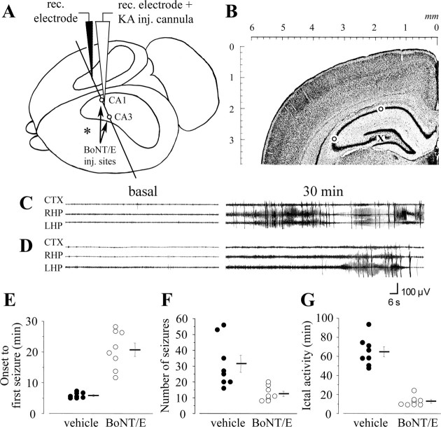
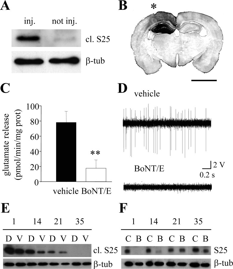
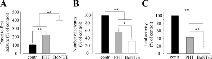
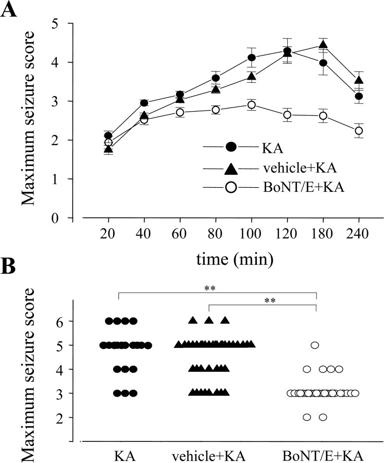
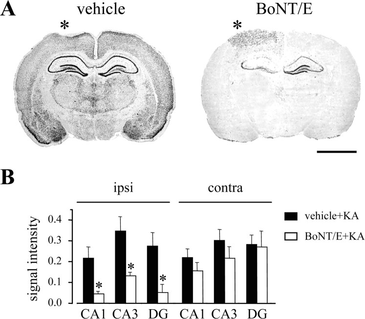
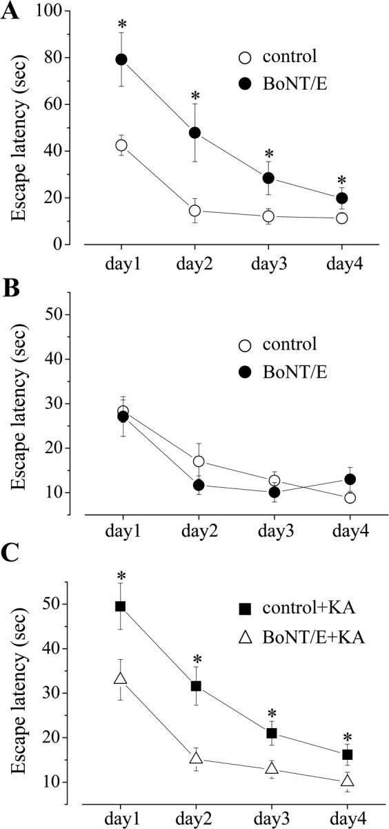
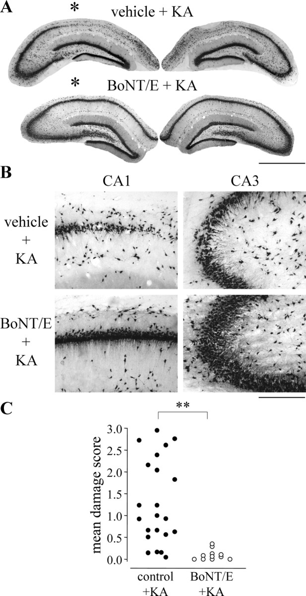
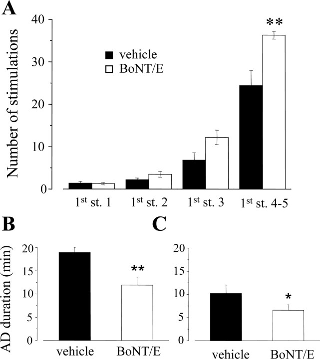
Experimental studies suggest that the delivery of antiepileptic agents into the seizure focus might be of potential utility for the treatment of focal-onset epilepsies. Botulinum neurotoxin E (BoNT/E) causes a prolonged inhibition of neurotransmitter release after its specific cleavage of the synaptic protein synaptosomal-associated protein of 25 kDa (SNAP-25). Here, we show that BoNT/E injected into the rat hippocampus inhibits glutamate release and blocks spike activity of pyramidal neurons. BoNT/E effects persist for at least 3 weeks, as determined by immunodetection of cleaved SNAP-25 and loss of intact SNAP-25. The delivery of BoNT/E to the rat hippocampus dramatically reduces both focal and generalized kainic acid-induced seizures as documented by behavioral and electrographic analysis. BoNT/E treatment also prevents neuronal loss and long-term cognitive deficits associated with kainic acid seizures. Moreover, BoNT/E-injected rats require 50% more electrical stimulations to reach stage 5 of kindling, thus indicating a delayed epileptogenesis. We conclude that BoNT/E delivery to the hippocampus is both antiictal and antiepileptogenic in experimental models of epilepsy.


Figures

Figure 2.
Figure 2.
Antiictal effect of BoNT/E on EEG seizures induced by intrahippocampal KA. A, Three-dimensional drawing of the rat brain illustrating the location of BoNT/E injection (inj.) sites (open circles) and the placement of KA injection cannula and recording (rec.) electrodes. Position of the cortical electrode is indicated by an asterisk. The plane of the coronal section shown in B, corresponding to stereotaxic coordinate AP -2.4 mm from bregma (see Materials and Methods), is also indicated. B, Nissl-stained coronal section through the hippocampus indicating the sites of BoNT/E injection (open circles) and the position of the KA infusion cannula glued to the recording electrode (X). C, D, EEG tracing depicting seizure activity induced by unilateral injection of 40 ng of KA into the dorsal hippocampus of rats that received 2 d before the convulsant either vehicle (C) or BoNT/E (D). Traces on the left represent baseline recordings in the parietal cortex (CTX), right hippocampus (RHP), and left hippocampus (LHP). Traces on the right depict typical seizure episodes. Time elapsed from the KA injection is indicated. E-G, Quantification of EEG seizures in control (black circles) and BoNT/E-injected (open circles) animals (n = 8 per group). A significant reduction in the number and duration of EEG seizures was consistently detected in BoNT/E-treated rats. The onset time to the first seizure, the total number of seizures, and the total time spent in ictal activity are reported in E-G, respectively. Each point indicates one animal, and horizontal bars represent mean values ± SE.
Figure 1.
Figure 1.
Characterization of BoNT/E effects in the hippocampus. A, Representative immunoblotting for cleaved SNAP-25 (cl. S25) on protein extracts from the BoNT/E-injected (inj.) and contralateral, noninjected (not inj.) hippocampus of a P36 rat, 1 d after BoNT/E injection. β-tub, β-Tubulin (internal standard). B, Forebrain coronal section showing the hippocampus of a P36 rat, 1 d after unilateral BoNT/E injection. Immunostaining for cleaved SNAP-25 (dark labeling) is mainly restricted to the injected hippocampus (indicated by an asterisk). Scale bar, 3 mm. C, Effects of BoNT/E on Ca2+-dependent, 35 mm K+-evoked glutamate over flow from hippocampal synaptosomes prepared 3 d after toxin injection. Release is inhibited by ∼80% in BoNT/E-injected hippocampi. Data are expressed as mean ± SE (n = 7 animals per group; **p < 0.01; t test). D, Representative recordings of spontaneous spike activity from CA1 in vehicle- and BoNT/E-injected animals. E, Immunoblotting for cleaved SNAP-25 (cl. S25) on protein extracts from hippocampi of BoNT/E-treated rats at different times after BoNT/E injection. D, Dorsal hippocampus; V, ventral hippocampus. F, Immunoblotting for the intact SNAP-25 (S25) on protein extracts from hippocampi of BoNT/E-treated (B) and age-matched control (C) rats at different times after BoNT/E injection.
Figure 3.
Figure 3.
Comparison of the antiictal effects of PHT (50 mg/kg, i.p.; n = 7 rats) and intrahippocampal BoNT/E (n = 8 rats). Drugs were given 60 min (PHT) or 2 d (BoNT/E) before intrahippocampal KA. Graphs report the onset time to first the seizure (A), the total number of seizures (B), and the total time spent in ictal activity (C) expressed as percentage of the respective control (contr) values. Control animals (n = 15) received intrahippocampal KA and the respective vehicles. *p < 0.05; **p < 0.01; one way ANOVA followed by Tukey test. Error bars represent SE.
Figure 4.
Figure 4.
BoNT/E prevents seizures induced by systemic KA. A, Progression of behavioral changes after systemic KA administration (8 mg/kg, i.p.) in the various treatment groups over a 4 h observation period (for details, see Materials and Methods). Data are mean seizure scores ± SE. B, Scatter plot reporting the maximum seizure score assigned to each experimental animal during a 4 h observation period after KA administration. The majority of the control animals reached status epilepticus (seizure stage 5), whereas only 1 of 30 BoNT/E-injected rats did so. **p < 0.01; one-way ANOVA followed by Dunn's test.
Figure 5.
Figure 5.
BoNT/E markedly decreases KA-induced c-fos mRNA expression in the brain. A, In situ hybridization analysis of c-fos mRNA in coronal brain sections at the level of the dorsal hippocampus from a vehicle- and a BoNT/E-injected rat, 2.5 h after systemic KA. The vehicle-injected rat reached status epilepticus (stage 5), whereas the BoNT/E-injected animal showed only preconvulsive behaviors (stage 3). Asterisks indicate the injected sides. Scale bar, 3 mm. B, Quantification of KA-induced c-fos mRNA expression in the CA1, CA3, and DG sectors of the hippocampus from vehicle- (black columns) and BoNT/E- (white columns) treated rats. Separate quantifications were made for the hippocampus ipsilateral (ipsi) and contralateral (contra) to the injected side. Signal intensity is expressed as integrated optical density ± SE (n = 5 animals per group; *p < 0.05; Student's t test).
Figure 6.
Figure 6.
BoNT/E prevents spatial learning deficits induced by KA. A, Acquisition of place learning in the Morris water maze (MWM) for rats injected with BoNT/E into the hippocampus at P35 and tested 3-7 d later (BoNT/E; n = 8; black circles). Age-matched control rats were injected intrahippocampally with vehicle (control; n = 8; open circles). Time required to find the submerged platform (escape latency) is indicated in seconds and plotted as mean values ± SE across days of testing. Performance of vehicle-treated rats is better than that of BoNT/E-treated rats (two-way ANOVA; p < 0.001). B, Acquisition of place learning in the MWM for rats injected with BoNT/E into the hippocampus at P35 and tested 5 weeks later (BoNT/E; n = 5; black circles). Performance of naive, age-matched rats (control; n = 10) is shown by open circles and is superimposable to that of BoNT/E-treated rats. No statistically significant difference is observed between the two groups (two-way ANOVA; p = 0.38). C, Acquisition of place learning in the MWM for rats injected with BoNT/E at P35 and injected with KA at P36 (BoNT/E+KA; n = 13; open triangles). Performance of control plus KA rats (n = 26) is shown by black squares and includes data from both naive (n = 7) and vehicle-injected (n = 19) rats treated with KA at P36 because these groups did not differ. Behavioral testing began for all rats 5 weeks after KA. Control plus KA rats were slower to learn the task and never reached the performance of the BoNT/E-injected group. The asterisks in A and C indicate p < 0.01 (two-way ANOVA followed by post hoc Tukey test).
Figure 7.
Figure 7.
BoNT/E prevents hippocampal neuronal loss induced by KA. A, Representative coronal sections through the dorsal hippocampus of P77 rats that received vehicle (top) or BoNT/E (bottom) at P35 and KA at P36. Sections were immunostained for the neuronal marker NeuN. Note preservation of hippocampal neurons in the BoNT/E-injected rat, whereas widespread neuronal loss is evident in the vehicle-injected animal. The injected side is indicated by an asterisk. B, Representative pictures showing details of CA1 and CA3 regions of the hippocampus from vehicle- and BoNT/E-injected rats. In the vehicle-treated rat, restricted areas of degeneration and reduced thickness of pyramidal cell layer (damage score 1; see Materials and Methods) are evident in CA3, whereas a marked loss of pyramidal neurons (damage score 2) is observed in CA1. Preservation of hippocampal tissue (no damage, score 0) is detected in both CA1 and CA3 regions of the BoNT/E-treated animal. Scale bars: A, 1.2 mm; B, 200 μm. C, Quantification of histological lesions in the dorsal hippocampus of control (control+KA; black circles; n = 22) and BoNT/E-injected (BoNT/E+KA; open circles; n = 11) rats treated with KA. The control plus KA group pools data from both naive and vehicle-injected rats because these two groups did not differ. The extent of the loss of hippocampal pyramidal neurons was determined for each animal as the mean damage score value in CA1 and CA3 sectors of both hemispheres (for details, see Materials and Methods). Each point represents one animal. **p < 0.01; Mann-Whitney rank-sum test.
Figure 8.
Figure 8.
BoNT/E delays hippocampal kindling rate. A, Behavioral progression of seizures during kindling in vehicle- (black columns) and BoNT/E- (white columns) injected rats. BoNT/E-treated animals required more stimulations to reach stages (st.) 4 and 5 of kindling. Data represent mean ± SE (**p < 0.01; two-way ANOVA followed by post hoc Tukey test). Behavioral stages are scored according to Racine's classification (Vezzani et al., 2002). B, C, Duration of primary (B) and secondary (C) afterdischarge during kindling in BoNT/E- and vehicle-injected rats. Cumulative AD was reckoned in each rat by adding together the durations of the single AD induced in the hippocampus during kindling stimulations. Data represent the mean ± SE of cumulative AD. *p < 0.05; **p < 0.01; Student's t test.

References

    1. Ashton AC, Dolly JO (1988) Characterization of the inhibitory action of botulinum neurotoxin type A on the release of several transmitters from rat cerebrocortical synaptosomes. J Neurochem 50: 1808-1816. - PubMed
    1. Ben-Ari Y (1985) Limbic seizure and brain damage produced by kainic acid: mechanisms and relevance to human temporal lobe epilepsy. Neuroscience 14: 375-403. - PubMed
    1. Bigalke H, Heller I, Bizzini B, Habermann E (1981) Tetanus toxin and botulinum A toxin inhibit release and uptake of various transmitters, as studied with particulate preparations from rat brain and spinal cord. Naunyn Schmiedebergs Arch Pharmacol 316: 244-251. - PubMed
    1. Bozzi Y, Vallone D, Borrelli E (2000) Neuroprotective role of dopamine against hippocampal cell death. J Neurosci 20: 8643-8649. - PMC - PubMed
    1. Caleo M, Medini P, von Bartheld CS, Maffei L (2003) Provision of brain-derived neurotrophic factor via anterograde transport from the eye preserves the physiological responses of axotomized geniculate neurons. J Neurosci 23: 287-296. - PMC - PubMed

Publication types

MeSH terms