. 2005 Feb 23;25(8):2002-9.
doi: 10.1523/JNEUROSCI.4474-04.2005.

Parkin mediates nonclassical, proteasomal-independent ubiquitination of synphilin-1: implications for Lewy body formation

Affiliations

Parkin mediates nonclassical, proteasomal-independent ubiquitination of synphilin-1: implications for Lewy body formation

Kah Leong Lim et al. J Neurosci. .

Abstract

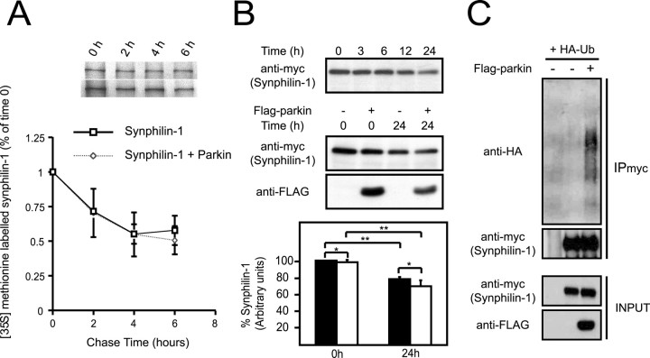
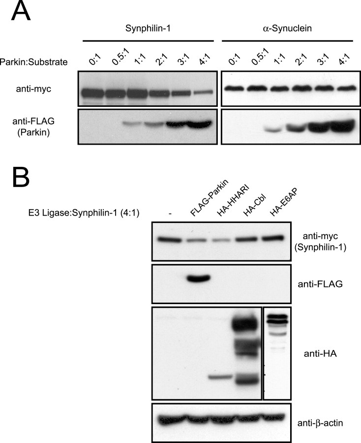
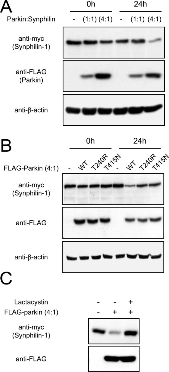
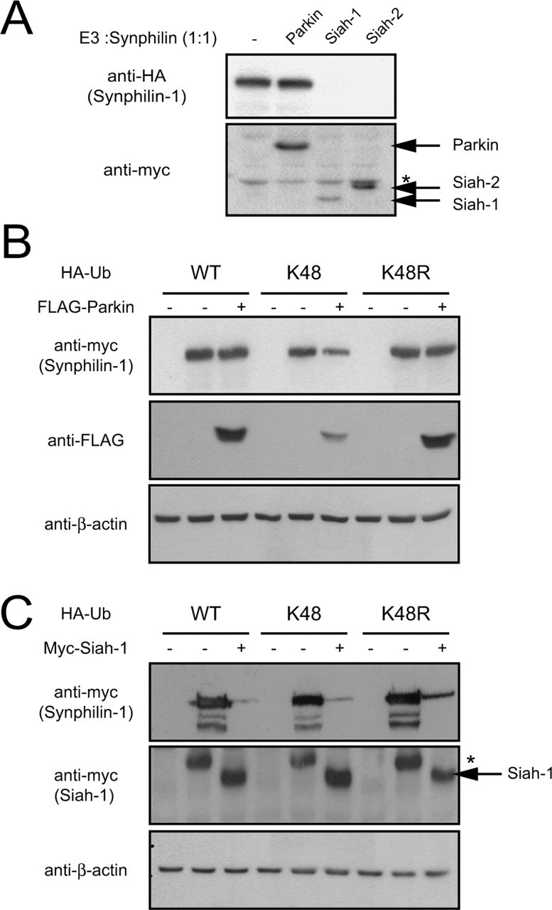
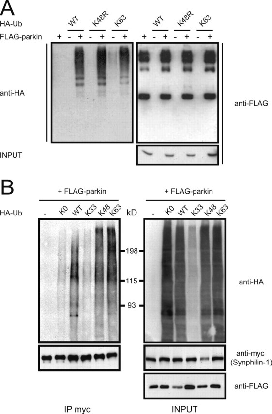
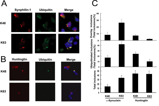
It is widely accepted that the familial Parkinson's disease (PD)-linked gene product, parkin, functions as a ubiquitin ligase involved in protein turnover via the ubiquitin-proteasome system. Substrates ubiquitinated by parkin are hence thought to be destined for proteasomal degradation. Because we demonstrated previously that parkin interacts with and ubiquitinates synphilin-1, we initially expected synphilin-1 degradation to be enhanced in the presence of parkin. Contrary to our expectation, we found that synphilin-1 is normally ubiquitinated by parkin in a nonclassical, proteasomal-independent manner that involves lysine 63 (K63)-linked polyubiquitin chain formation. Parkin-mediated degradation of synphilin-1 occurs appreciably only at an unusually high parkin to synphilin-1 expression ratio or when primed for lysine 48 (K48)-linked ubiquitination. In addition we found that parkin-mediated ubiquitination of proteins within Lewy-body-like inclusions formed by the coexpression of synphilin-1, alpha-synuclein, and parkin occurs predominantly via K63 linkages and that the formation of these inclusions is enhanced by K63-linked ubiquitination. Our results suggest that parkin is a dual-function ubiquitin ligase and that K63-linked ubiquitination of synphilin-1 by parkin may be involved in the formation of Lewy body inclusions associated with PD.


Figures

Figure 1.
Figure 1.
Effect of parkin on synphilin-1 turnover. A, Pulsechase analysis of the turnover rate of myc-tagged synphilin-1 in HEK 293 cells in the absence or presence of FLAG-tagged parkin. Data from three independent experiments are expressed as means ± SEM. B, Top panel, Anti-myc immunoblot of equal amount of total cell lysates prepared from HEK 293 cells transfected with myc-tagged synphilin-1 and treated with puromycin (100 μm) for different durations as indicated. Middle panel, Anti-myc and anti-FLAG immunoblots of equal amount of total cell lysates prepared from HEK 293 cells transfected with myc-tagged synphilin-1 without or with an equivalent amount of FLAG-tagged parkin and treated with puromycin (100 μm) for 24 h. The anti-myc immunoblots from three independent experiments were used to derive the relative densitometric units of synphilin-1, which is presented as a histogram in the bottom panel. Closed and opened bars represent units of synphilin-1 in the absence and presence of parkin respectively (*p > 0.1; **p < 0.05; Student's t test). C, Anti-HA and anti-myc immunoblots of synphilin immunoprecipitates (IPmyc) prepared from HEK 293 cells transfected with myc-tagged synphilin-1 in the presence of HA-tagged wild-type ubiquitin without or with FLAG-tagged parkin. Lysates prepared from these variously transfected cells were also subjected to anti-myc or anti-FLAG immunoblotting to show their expression (INPUT). These experiments were replicated at least three times.
Figure 2.
Figure 2.
Dose-dependent effect of parkin on synphilin-1 steady-state levels. A, Anti-myc and anti-FLAG immunoblots of total cell lysates prepared from HEK 293 cells transfected with myc-tagged synphilin-1 (left panels) or myc-tagged α-synuclein (right panels) in the absence or presence of increasing relative amounts of FLAG-tagged parkin, as indicated. B, Anti-myc, anti-FLAG, and anti-HA immunoblots of total cell lysates prepared from HEK 293 cells transfected with myc-tagged synphilin-1 alone or with various FLAG-tagged or HA-tagged E3 ligases (as indicated) at an E3 ligase/synphilin-1 cDNA cotransfection ratio of 4:1. Equal loading of the different cell lysates was verified by anti-β-actin immunoblotting. These experiments were replicated at least three times.
Figure 3.
Figure 3.
Degradation of synphilin-1 by parkin is dependent on their relative expression levels and on parkin catalytic competency. A, Anti-myc and anti-FLAG immunoblots of total cell lysates prepared from HEK 293 cells transfected with myc-tagged synphilin-1 without or with FLAG-tagged parkin at different cotransfection ratios (as indicated) and treated 24 h later with puromycin (100 μm) for 24 h. B, Anti-myc and anti-FLAG immunoblots of total cell lysates prepared from HEK 293 cells transfected with myc-tagged synphilin-1 without or with FLAG-tagged, wild-type parkin, or familial-PD parkin mutants, at a parkin/synphilin-1 cDNA cotransfection ratio of 4:1 and treated with puromycin (100 μm) for 24 h. In both cases, equal loading of the different cell lysates was verified by anti-β-actin immunoblotting. C, HEK 293 cells transfected with the indicated combinations of myc-tagged synphilin-1 and FLAG-tagged parkin were left untreated or treated with 5 μm clasto-lactacytstin β-lactone (lactacystin) for 16 h. Lysates prepared from these cells were immunoblotted with anti-myc or anti-FLAG as indicated. These experiments were duplicated with similar results.
Figure 4.
Figure 4.
Parkin is not an efficient degradation-associated E3 ligase. A, Anti-HA and anti-myc immunoblots of total cell lysates prepared from HEK 293 cells transfected with HA-tagged synphilin-1 alone or with myc-tagged parkin, siah-1, or siah-2 at 1:1 cotransfection ratio. Asterisk indicates nonspecific band. B, Anti-myc and anti-FLAG immunoblots of total cell lysates prepared from HEK 293 cells cotransfected with the indicated combinations of myc-tagged synphilin-1, FLAG-tagged parkin, and various HA-tagged ubiquitin species. C, Anti-myc immunoblots of total cell lysates prepared from HEK 293 cells cotransfected with the indicated combinations of myc-tagged synphilin-1, myc-tagged siah-1, and various HA-tagged ubiquitin species. Asterisk indicates nonspecific band. For both B and C, equal loading of the different cell lysates was verified by anti-β-actin immunoblotting. These experiments were duplicated with similar results.
Figure 5.
Figure 5.
Parkin mediates K63-linked polyubiquitination of synphilin-1. A, A portion of lysates prepared from HEK 293 cells transfected with FLAG-tagged parkin alone or with various HA-tagged ubiquitin species were subjected to anti-FLAG immunoprecipitation followed by anti-HA immunoblotting (left panel). The blot was stripped and reprobed with anti-FLAG to show the amounts of immunoprecipitated parkin (top right panel). Lysates prepared from these variously transfected cells (INPUT) were also subjected to anti-FLAG immunoblotting to show FLAG-parkin expression levels (bottom right panel). B, A portion of lysates prepared from HEK 293 cells transfected with the indicated combinations of myc-tagged synphilin-1, FLAG-tagged parkin, and various HA-tagged ubiquitin species were subjected to anti-myc immunoprecipitation followed by anti-HA immunoblotting (top left panel). The blot was stripped and reprobed with anti-myc (bottom left panel) to illustrate that relatively equal amounts of synphilin were immunoprecipitated. The remaining portion of the transfected cell lysates was immunoblotted with anti-HA (top right panel) and anti-myc (bottom right panel) to show their respective expression levels. These experiments were replicated at least three times.
Figure 6.
Figure 6.
Parkin mediates predominantly K-63-linked ubiquitination of cytosolic inclusions formed by coexpression of synphilin-1 and α-synuclein. A, Coexpression of α-synuclein, myc-tagged synphilin-1, FLAG-tagged parkin, and either HA-tagged K48 ubiquitin or K63 ubiquitin results in the formation of myc-synphilin-1-positive inclusions (red). Positive anti-HA staining of the inclusion bodies is observed only in transfections containing HA-tagged K63 ubiquitin (green). The merged pictures show colocalization of the anti-myc and anti-HA staining (yellow). B, Coexpression of mutant huntingtin and either HA-tagged K-48 ubiquitin or HA-tagged K-63 ubiquitin results in the formation of huntingtin-positive inclusions (red). Positive anti-HA staining of the inclusion bodies is observed only in transfections containing HA-tagged K-48 ubiquitin (green). The merged pictures show colocalization of the anti-huntingtin and anti-HA staining (yellow). C, Histograms showing quantitative data of inclusion bodies in SH-SY5Y cells cotransfected with the various construct as described in A and B. These experiments were replicated at least three times.

References

    1. Ardley HC, Tan NG, Rose SA, Markham AF, Robinson PA (2001) Features of the parkin/ariadne-like ubiquitin ligase, HHARI, that regulate its interaction with the ubiquitin-conjugating enzyme, Ubch7. J Biol Chem 276: 19640-19647. - PubMed
    1. Arnason T, Ellison MJ (1994) Stress resistance in Saccharomyces cerevisiae is strongly correlated with assembly of a novel type of multiubiquitin chain. Mol Cell Biol 14: 7876-7883. - PMC - PubMed
    1. Chau V, Tobias JW, Bachmair A, Marriott D, Ecker DJ, Gonda DK, Varshavsky A (1989) A multiubiquitin chain is confined to specific lysine in a targeted short-lived protein. Science 243: 1576-1583. - PubMed
    1. Choi P, Snyder H, Petrucelli L, Theisler C, Chong M, Zhang Y, Lim K, Chung KK, Kehoe K, D'Adamio L, Lee JM, Cochran E, Bowser R, Dawson TM, Wolozin B (2003) SEPT5_v2 is a parkin-binding protein. Brain Res Mol Brain Res 117: 179-189. - PubMed
    1. Chung KK, Dawson VL, Dawson TM (2001a) The role of the ubiquitin-proteasomal pathway in Parkinson's disease and other neurodegenerative disorders. Trends Neurosci 24: S7-14. - PubMed

Publication types

MeSH terms