. 2005 Mar;49(3):1127-34.
doi: 10.1128/AAC.49.3.1127-1134.2005.

Telavancin, a multifunctional lipoglycopeptide, disrupts both cell wall synthesis and cell membrane integrity in methicillin-resistant Staphylococcus aureus

Affiliations

Telavancin, a multifunctional lipoglycopeptide, disrupts both cell wall synthesis and cell membrane integrity in methicillin-resistant Staphylococcus aureus

Deborah L Higgins et al. Antimicrob Agents Chemother. 2005 Mar.

Abstract

The emergence and spread of multidrug-resistant gram-positive bacteria represent a serious clinical problem. Telavancin is a novel lipoglycopeptide antibiotic that possesses rapid in vitro bactericidal activity against a broad spectrum of clinically relevant gram-positive pathogens. Here we demonstrate that telavancin's antibacterial activity derives from at least two mechanisms. As observed with vancomycin, telavancin inhibited late-stage peptidoglycan biosynthesis in a substrate-dependent fashion and bound the cell wall, as it did the lipid II surrogate tripeptide N,N'-diacetyl-L-lysinyl-D-alanyl-D-alanine, with high affinity. Telavancin also perturbed bacterial cell membrane potential and permeability. In methicillin-resistant Staphylococcus aureus, telavancin caused rapid, concentration-dependent depolarization of the plasma membrane, increases in permeability, and leakage of cellular ATP and K(+). The timing of these changes correlated with rapid , concentration-dependent loss of bacterial viability, suggesting that the early bactericidal activity of telavancin results from dissipation of cell membrane potential and an increase in membrane permeability. Binding and cell fractionation studies provided direct evidence for an interaction of telavancin with the bacterial cell membrane; stronger binding interactions were observed with the bacterial cell wall and cell membrane relative to vancomycin. We suggest that this multifunctional mechanism of action confers advantageous antibacterial properties.


Figures

FIG. 1.
FIG. 1.
Inhibition of macromolecular synthesis in MRSA 33591 by telavancin, THRX-881620, and vancomycin. The percentage of inhibition of RNA (▪), fatty acid (▴), peptidoglycan (▾), and protein (⧫) and the percentage of killing (•) after incubation for 10 min are plotted versus drug concentration. Each point is the mean ± standard deviation of 3 estimates. The MICs of telavancin (A), THRX-881620 (B), and vancomycin (C) were 0.3, 8, and 0.7 μM, respectively. The IC50s for peptidoglycan synthesis inhibition determined from these concentration-inhibition curves were 0.14, 5.8, and 2.0 μM for telavancin, THRX-881620, and vancomycin, respectively.
FIG. 2.
FIG. 2.
Inhibition of peptidoglycan synthesis in MRSA 33591 cells. The percentages of peptidoglycan synthesis inhibited by various concentrations of telavancin (circles), THRX-881620 (squares), and vancomycin (triangles) in the absence (open symbols) and presence (closed symbols) of 0.5 mM dKAA are plotted. Each data point is the mean ± standard deviation of 3 estimates. The IC50s determined from these concentration-inhibition curves in the absence and presence of dKAA are 0.14 and 1.4 μM for telavancin, 3.2 and 3.3 μM for THRX-881620, and 4.8 and >100 μM for vancomycin.
FIG. 3.
FIG. 3.
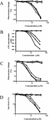
Correlation between change in membrane potential (A), ATP leakage (B), permeability (C), and cell viability (D) at various times after antibiotic addition. •, 15 min; ▪, 30 min; ▴, 60 min; closed symbols, telavancin; open symbols, vancomycin. Representative curves are shown from single experiments that were reproduced in at least three independent determinations.
FIG. 4.
FIG. 4.
Antagonism of telavancin-induced membrane permeability by dKAA. Membrane permeability was induced by 8 μM telavancin in the absence (•) or presence (added 10 min prior to telavancin) of the following concentrations of dKAA: 31.25 μM (▴), 62.5 mM (▵), 125 μΜ (▾), 250 μΜ (▿), 500 μΜ (▪). There was also a permeability control with no telavancin and no dKAA (○). Representative curves are shown from a single experiment that was repeated three times.

References

    1. Allen, N. E., J. N. Hobbs, Jr., and T. I. Nicas. 1996. Inhibition of peptidoglycan biosynthesis in vancomycin-susceptible and -resistant bacteria by a semisynthetic glycopeptide antibiotic. Antimicrob. Agents Chemother. 40:2356-2362. - PMC - PubMed
    1. Allen, N. E., D. L. LeTourneau, and J. N. Hobbs, Jr. 1997. Molecular interactions of a semisynthetic glycopeptide antibiotic with d-alanyl-d-alanine and d-alanyl-d-lactate residues. Antimicrob. Agents Chemother. 41:66-71. - PMC - PubMed
    1. Allen, N. E., D. L. LeTourneau, and J. N. Hobbs, Jr. 1997. The role of hydrophobic side chains as determinants of antibacterial activity of semisynthetic glycopeptide antibiotics. J. Antibiot. (Tokyo) 50:677-684. - PubMed
    1. Allen, N. E., D. L. LeTourneau, J. N. Hobbs, Jr., and R. C. Thompson. 2002. Hexapeptide derivatives of glycopeptide antibiotics: tools for mechanism of action studies. Antimicrob. Agents Chemother. 46:2344-2348. - PMC - PubMed
    1. Allen, N. E., and T. I. Nicas. 2003. Mechanism of action of oritavancin and related glycopeptide antibiotics. FEMS Microbiol. Rev. 26:511-532. - PubMed

MeSH terms