

Regional variation of intracortical porosity in the midshaft of the human femur: age and sex differences
- PMID: 15730477
- PMCID: PMC1571459
- DOI: 10.1111/j.1469-7580.2005.00384.x
Regional variation of intracortical porosity in the midshaft of the human femur: age and sex differences
Abstract
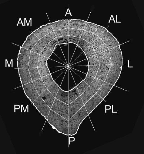
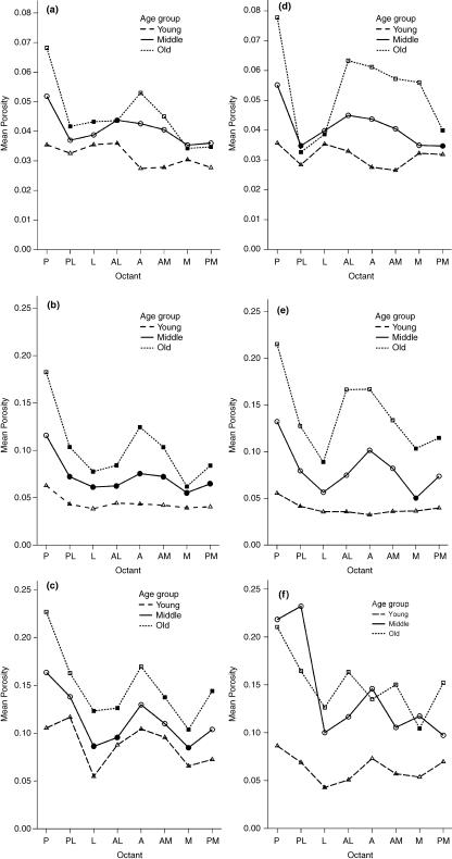
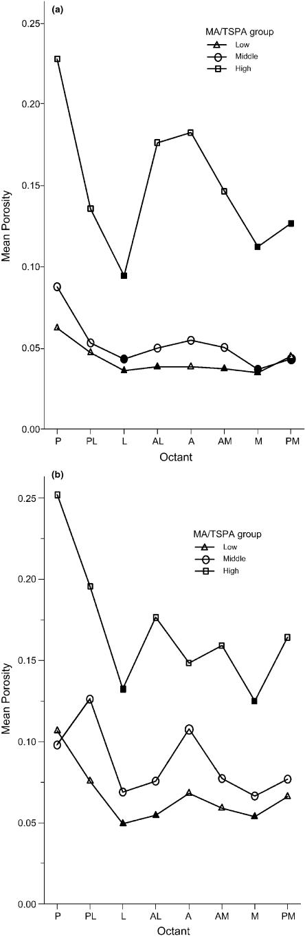
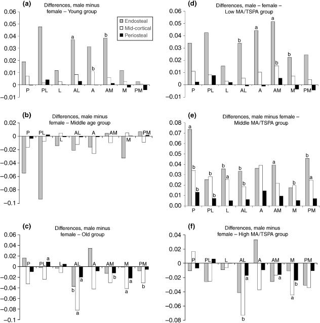
This study investigated age and sex differences in patterns of porosity distribution in the midshaft of the human femur. Cross-sections were obtained from 168 individuals from a modern Australian population. The sample comprised 73 females and 95 males, aged between 20 and 97 years. Microradiographs were made of 100-microm sections and pore and bone areas were determined using image processing software. Initially the sample was divided by age: young (20-44 years), middle (45-64 years) and old (65+ years), but it was found that analysis on the basis of the ratio of medullary area to total subperiosteal area gave clearer results. The cortex was divided into three rings radially and into octants circumferentially and the porosity of each segment was calculated. Results showed that a pattern with raised porosity in the posterior and anterolateral regions, and with greater porosity in the inner parts of the cortex, becomes more pronounced with age. In males this pattern develops steadily; in females there are much greater differences between the middle and older groups than earlier in life. The patterns observed are consistent with progressive bone loss occurring along a neutral axis of the cortex where bending stress is lowest and the mechanical advantage of the bone is least.
Figures
References
-
- Ahlqvist M, Damsten O. A modification of Kerley's method for the microscopic determination of age in human bone. J. Forensic Sci. 1969;14:205–212. - PubMed
-
- Atkinson PJ. Changes in resorption spaces in femoral cortical bone with age. J. Path. Bacteriol. 1965;89:173–178. - PubMed
-
- Baer MJ. Growth and Maturation. an Introduction to Physical Development. Cambridge, MA: Howard A Doyle; 1977.
-
- Bell KL, Loveridge N, Power J, et al. Structure of the femoral neck in hip fracture: Cortical bone loss in the inferoanterior to superoposterior axis. J. Bone Miner Res. 1999a;14:111–119. - PubMed
-
- Bell KL, Loveridge N, Power J, Garrahan N, Meggitt BF, Reeve J. Regional differences in cortical porosity in the fractured femoral neck. Bone. 1999b;24:57–64. - PubMed
MeSH terms
LinkOut - more resources
Full Text Sources
Medical
