

Relationships among microstructural properties of bone at the human midshaft femur
- PMID: 15730478
- PMCID: PMC1571464
- DOI: 10.1111/j.1469-7580.2005.00385.x
Relationships among microstructural properties of bone at the human midshaft femur
Abstract
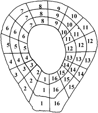
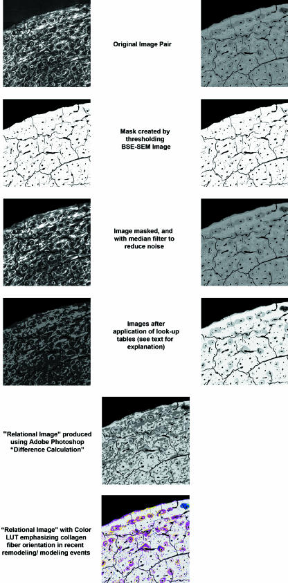
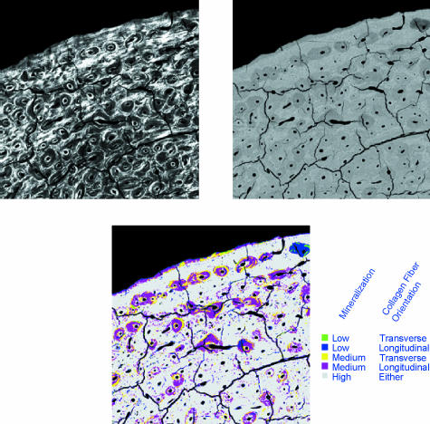
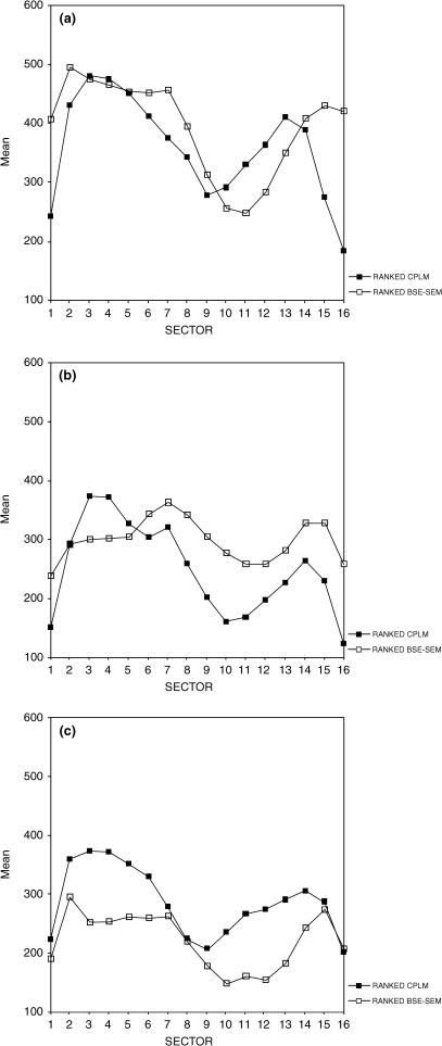
Mineralization density and collagen fibre orientation are two aspects of a bone's microstructural organization that influence its mechanical properties. Previous studies by our group have demonstrated a distinctly non-random, though highly variable, spatial distribution of these two variables in the human femoral cortex. In this study of 37 specimens, these variables are examined relative to one another in order to determine whether regions of bone demonstrating higher or lower mineralization density also demonstrate a prevalence of either transversely or longitudinally oriented collagen fibres. An analysis of rank-transformed collagen fibre orientation (as determined by circularly polarized light) and mineralization density (as determined by backscattered electron microscopy) data sets demonstrated that areas of low mineralization density (predominantly in the anterior-lateral cortex) tended to correspond to regions of higher proportions of longitudinally oriented collagen fibres. Conversely, areas of higher mineralization density (postero-medially) tended to correspond to regions of higher proportions of transversely oriented collagen fibres. High variability in the sample led to generally low correlations between the two data sets, however. A second analysis focused only on the orientation of collagen fibres within poorly mineralized bone (representing bone that was newly formed). This analysis demonstrated a lower proportion of transverse collagen fibres in newly formed bone with age, along with some significant regional differences in the prevalence of collagen fibres of either orientation. Again high variability characterized the sample. These results are discussed relative to the hypothesized forces experienced at the midshaft femur.
Figures
Similar articles
-
Intrapopulation variability in mineralization density at the human femoral mid-shaft.J Anat. 2003 Aug;203(2):243-55. doi: 10.1046/j.1469-7580.2003.00212.x. J Anat. 2003. PMID: 12924824 Free PMC article.
-
Preferred collagen fiber orientation in the human mid-shaft femur.Anat Rec A Discov Mol Cell Evol Biol. 2003 May;272(1):434-45. doi: 10.1002/ar.a.10055. Anat Rec A Discov Mol Cell Evol Biol. 2003. PMID: 12704701
-
Peri-implant bone organization under immediate loading conditions: collagen fiber orientation and mineral density analyses in the minipig model.Clin Implant Dent Relat Res. 2009 Mar;11(1):41-51. doi: 10.1111/j.1708-8208.2008.00086.x. Epub 2008 Jul 23. Clin Implant Dent Relat Res. 2009. PMID: 18657155
-
Circularly polarized light standards for investigations of collagen fiber orientation in bone.Anat Rec B New Anat. 2003 Sep;274(1):157-68. doi: 10.1002/ar.b.10031. Anat Rec B New Anat. 2003. PMID: 12964206 Review.
-
Natural factors that affect the shape and strength of the aging human femur.Clin Orthop Relat Res. 1992 Jan;(274):194-201. Clin Orthop Relat Res. 1992. PMID: 1729003 Review.
Cited by
-
Revealing Developmental Transitions in Perinatal and Infant Individuals Through Microanatomical Analysis.Am J Hum Biol. 2025 Jul;37(7):e70101. doi: 10.1002/ajhb.70101. Am J Hum Biol. 2025. PMID: 40658068 Free PMC article.
-
A Review of Histological Techniques for Differentiating Human Bone from Animal Bone.Methods Protoc. 2024 Jun 30;7(4):51. doi: 10.3390/mps7040051. Methods Protoc. 2024. PMID: 39051265 Free PMC article. Review.
-
Alendronate treatment promotes bone formation with a less anisotropic microstructure during intramembranous ossification in rats.J Bone Miner Metab. 2008;26(1):24-33. doi: 10.1007/s00774-007-0782-8. Epub 2008 Jan 10. J Bone Miner Metab. 2008. PMID: 18095060
-
Spatial variation in osteon population density at the human femoral midshaft: histomorphometric adaptations to habitual load environment.J Anat. 2016 May;228(5):733-45. doi: 10.1111/joa.12433. Epub 2015 Dec 28. J Anat. 2016. PMID: 26708961 Free PMC article.
-
Hyperlipidemia affects multiscale structure and strength of murine femur.J Biomech. 2014 Jul 18;47(10):2436-43. doi: 10.1016/j.jbiomech.2014.04.006. Epub 2014 Apr 16. J Biomech. 2014. PMID: 24795172 Free PMC article.
References
-
- Amprino R, Bairati A. Processi di ricostruzione e di riassorbimento nella sostanza compatta delle ossa dellúomo. Z. Zellforsch. Mikrosk. 1936;24:439–511.
-
- Amtmann VE. Mechanical stress functional adaptation and the variation structure of the human femur diaphysis. Erg. Anat. Und. Ent. 1971;44:1–87. - PubMed
-
- Ascenzi A, Bonucci E. The tensile properties of single osteons. Anat. Rec. 1967;158:375–386. - PubMed
-
- Ascenzi A, Bonucci E. The compressive properties of single osteons. Anat. Rec. 1968;161:377–392. - PubMed
-
- Bertelsen PK, Clement JG, Thomas CDL. A morphometric study of the cortex of the human femur from early childhood to advanced old age. Forensic Sci. Int. 1995;74:63–77. - PubMed
Publication types
MeSH terms
Substances
LinkOut - more resources
Full Text Sources
Medical
