

Effects of glial cell line-derived neurotrophic factor on isolated developing mouse Sertoli cells in vitro
- PMID: 15730482
- PMCID: PMC1571465
- DOI: 10.1111/j.1469-7580.2005.00373.x
Effects of glial cell line-derived neurotrophic factor on isolated developing mouse Sertoli cells in vitro
Abstract
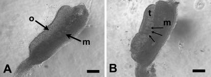
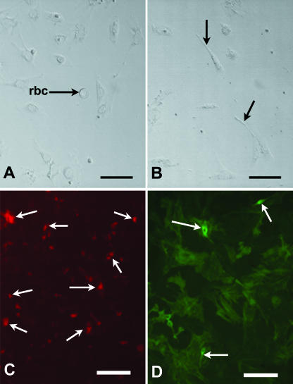
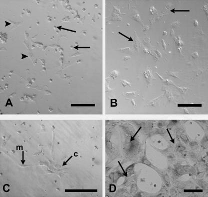
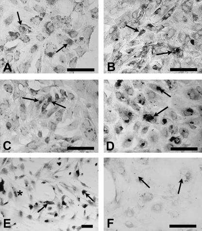
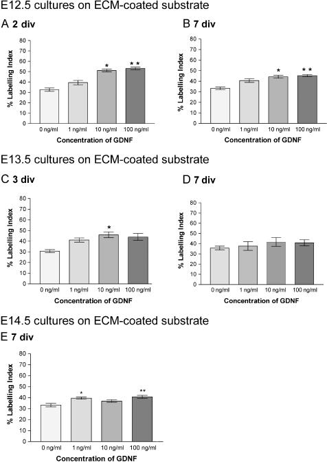
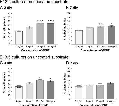
Cell proliferation is a key factor in sex determination where a size increase relative to the XX gonad is one of the first signs of testis differentiation. Moreover, proliferation of Sertoli cells during development is important in building up the stock of supporting cells necessary for subsequent successful fertility. Because proliferation is such an essential part of testis development, the hypothesis under long-term investigation is that it is under fail-safe control by multiple alternative growth factors. This study was undertaken to investigate the role of glial cell-derived neurotrophic factor (GDNF) on developing mouse Sertoli cells in vitro. Sertoli cells, isolated from mouse embryos at three stages of testis development, were maintained for 2-7 days in vitro (div) in the presence or absence of GDNF at 1, 10 and 100 ng mL(-1). Overall the presence of extracellular matrix gel had little effect on proliferative activity, but encouraged expression of the epithelial phenotype. A statistically significant difference in proliferation, assessed by immunocytochemical staining for proliferating cell nuclear antigen, was seen with GDNF at embryonic day (E)12.5 after 2 div (at both 10 and 100 ng mL(-1), P < 0.001) and 7 div (at both 10 and 100 ng mL(-1), P < 0.05); at E13.5 after 3 div (at both 10 and 100 ng mL(-1), P < 0.05) and at E14.5 after 7 div (100 ng mL(-1), P < 0.01), compared with controls cultured without growth factor. In conclusion, GDNF stimulates mitosis throughout this critical developmental window. The in vitro approach used here is a useful adjunct to the knockout mouse model and has been applied to show that GDNF exerts a proliferative effect on developing mouse Sertoli cells.
Figures
Similar articles
-
Glial cell line-derived neurotropic factor stimulates sertoli cell proliferation in the early postnatal period of rat testis development.Endocrinology. 1999 Aug;140(8):3416-21. doi: 10.1210/endo.140.8.6922. Endocrinology. 1999. PMID: 10433195
-
Effects of FGF9 on embryonic Sertoli cell proliferation and testicular cord formation in the mouse.Int J Dev Biol. 2004 Sep;48(7):637-43. doi: 10.1387/ijdb.031778lw. Int J Dev Biol. 2004. PMID: 15470636
-
Effects of glial cell line-derived neurotrophic factor, fibroblast growth factor 2 and epidermal growth factor on proliferation and the expression of some genes in buffalo (Bubalus bubalis) spermatogonial cells.Reprod Fertil Dev. 2013;25(8):1149-57. doi: 10.1071/RD12330. Reprod Fertil Dev. 2013. PMID: 23171731
-
Regulation of GDNF expression in Sertoli cells.Reproduction. 2019 Mar;157(3):R95-R107. doi: 10.1530/REP-18-0239. Reproduction. 2019. PMID: 30620720 Free PMC article. Review.
-
The Regulation of Spermatogonial Stem Cells in an Adult Testis by Glial Cell Line-Derived Neurotrophic Factor.Front Endocrinol (Lausanne). 2022 Jun 3;13:896390. doi: 10.3389/fendo.2022.896390. eCollection 2022. Front Endocrinol (Lausanne). 2022. PMID: 35721702 Free PMC article. Review.
Cited by
-
Identification of candidate gonadal sex differentiation genes in the chicken embryo using RNA-seq.BMC Genomics. 2015 Sep 16;16(1):704. doi: 10.1186/s12864-015-1886-5. BMC Genomics. 2015. PMID: 26377738 Free PMC article.
-
GDNF enhances HGF-induced tubulogenesis and organization of Sertoli cell.J Assist Reprod Genet. 2025 Jun;42(6):2083-2098. doi: 10.1007/s10815-025-03493-7. Epub 2025 May 22. J Assist Reprod Genet. 2025. PMID: 40402398 Free PMC article.
-
Gdnf Acts as a Germ Cell-Derived Growth Factor and Regulates the Zebrafish Germ Stem Cell Niche in Autocrine- and Paracrine-Dependent Manners.Cells. 2022 Apr 11;11(8):1295. doi: 10.3390/cells11081295. Cells. 2022. PMID: 35455974 Free PMC article.
-
Glial-derived neurotrophic factor promotes ovarian primordial follicle development and cell-cell interactions during folliculogenesis.Reproduction. 2008 May;135(5):671-82. doi: 10.1530/REP-07-0405. Epub 2008 Feb 27. Reproduction. 2008. PMID: 18304989 Free PMC article.
-
TGF-β superfamily: how does it regulate testis development.Mol Biol Rep. 2012 Apr;39(4):4727-41. doi: 10.1007/s11033-011-1265-5. Epub 2011 Sep 27. Mol Biol Rep. 2012. PMID: 21947950 Review.
References
-
- Campagnolo L, Russo MA, Puglianiello A, Favale A, Siracusa G. Mesenchymal precursors of peritubular smooth muscle cells of the mouse testis can be identified by the presence of the p75 neurotrophin receptor. Biol. Reprod. 2001;64:464–472. - PubMed
-
- Colvin J, Green RP, Schmahl J, Capel B, Ornitz D. Male-to-female sex reversal in mice lacking fibroblast growth factor 9. Cell. 2001;104:875–889. - PubMed
-
- Cupp AS, Tessarollo L, Skinner MK. Testis developmental phenotypes in neurotrophin receptor trkA and trkC null mutations: role in formation of seminiferous cords and germ cell survival. Biol. Reprod. 2002;66:1838–1845. - PubMed
-
- Cupp AS, Uzumcu M, Skinner MK. Chemotactic role of neurotropin-3 in the embryonic testis that facilitates male sex determination. Biol. Reprod. 2003;68:2033–2037. - PubMed
-
- Golden JP, DeMaro JA, Osborne PA, Milbrandt J, Johnson EM., Jr Expression of neurturin, GDNF and GDNF family-receptor mRNA in the developing and mature mouse. Exp. Neurol. 1999;158:504–528. - PubMed
Publication types
MeSH terms
Substances
Grants and funding
LinkOut - more resources
Full Text Sources
