. 2005 Mar;73(3):1584-9.
doi: 10.1128/IAI.73.3.1584-1589.2005.

Rapid loss of motility of Helicobacter pylori in the gastric lumen in vivo

Affiliations
Free PMC article

Rapid loss of motility of Helicobacter pylori in the gastric lumen in vivo

Sören Schreiber et al. Infect Immun. 2005 Mar.
Free PMC article

Abstract

The human pathogen Helicobacter pylori has infected more than half of the world's population. Nevertheless, the first step of infection, the acute colonization of the gastric mucus, is poorly understood. For successful colonization, H. pylori must retain active motility in the gastric lumen until it reaches the safety of the mucus layer. To identify the factors determining the acute colonization, we inserted bacteria into the stomach of anesthetized Mongolian gerbils. We adjusted the gastric juice to defined pH values of between 2.0 and 6.0 by using an autotitrator. Despite the fact that Helicobacter spp. are known to survive low pH values for a certain time in vitro, the length of time that H. pylori persisted under the assay conditions within the gastric juice in vivo was remarkably shorter. In the anesthetized animal we found H. pylori to be irreversibly immotile in less than 1 min at lumen pH values of 2 and 3. At pH 4 motility was lost after 2 min. However, the period of motility increased to more than 15 min at pH 6. Blocking pepsins in the gastric lumen in vivo by using pepstatin significantly increased the period of motility. It was possible to simulate the rapid in vivo immotilization in vitro by adding pepsins. We conclude that pepsin limits the persistence of H. pylori in the gastric chymus to only a few minutes by rapidly inhibiting active motility. It is therefore likely that this short period of resistance in the gastric lumen is one of the most critical phases of Helicobacter infection.


Figures

FIG. 1.
FIG. 1.
Setup for the measurement of bacterial motility in vivo. The stomach of an anesthetized gerbil was lifted onto a micromanipulator. Through a small incision a combination micro pH electrode, a stirrer, and the tube of the autotitrator were inserted into the antrum region of the stomach. Then, 50 μl of the bacterial suspension were added to 0.5 to 1 ml of gastric juice. A gas flow of 5% O2, 10% CO2, and 85% N2 was applied over the opened stomach to maintain a microaerobic climate. The pH electrode continuously measured the luminal pH and the autotitrator held the pH at the test value by applying 0.25 N HCl and 100 mM urea. The stirrer ensured efficient mixing of the luminal content. During the experiment the stomach wall was covered by wet cotton wool.
FIG. 2.
FIG. 2.
Setup for the measurement of bacterial motility in vitro. To test the bacterial motility in vitro, 100 μl of bacterial suspension was incubated in a reaction vial, which was inserted into a chamber of water maintained at 37°C. The pH in the suspension was measured by a combination pH-electrode and was automatically titrated by adding a solution of 0.25 N HCl and 100 mM urea. Another small tube was inserted into the reaction vial, which applied a gas flow of 5% O2, 10% CO2, and 85% N2. The small gas bubbles immediately surfaced and thus mixed the contents of the vial (gas lift). The remaining space in the chamber was filled with the same gas mixture, which subsequently left the chamber through a filtered outlet.
FIG. 3.
FIG. 3.
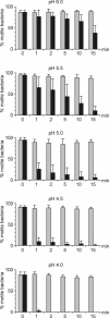
Motility of H. pylori at pH 6.0, 5.5, 5.0, 4.5, and 4.0 in vitro (░⃞) and in the stomach lumen of anesthetized Mongolian gerbils (▪). Bacteria were sampled after 1, 2, 5, 10, and 15 min, and their motility assessed microscopically. The number of motile bacteria in each sample was given as a percentage. The bacterial motility under the in vitro conditions remained unaffected for the entire experiment. Five animals and five bacterial suspensions are shown in each graph (mean percentage of motile bacteria ± the SD). All values (with the exception of the pH 6 first and second minute and the pH 5.5 first minute) from 1 to 15 min of exposure time at all pH values in vivo were significantly different from the in vitro values (Student t test, P < 0.05).
FIG. 4.
FIG. 4.
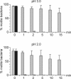
Motility of H. pylori at pH 3.0 and 2.0 in vitro (░⃞) and in the stomach lumen of the anesthetized Mongolian gerbil (▪). At these low pH values, the motility already decreases in the in vitro samples over the time period of the experiment. The bacterial motility under in vivo conditions was lost within the first minute. Five animals and five bacterial suspensions are shown in each graph (mean percentage of motile bacteria ± the SD). All values from 1 to 15 min of exposure time at all pH values in vivo were significantly different from the in vitro values (Student t test, P < 0.05).
FIG. 5.
FIG. 5.
Loss of motility of H. pylori in the stomach lumen of the Mongolian gerbil at pH 5.0, 4.5, 4.0, 3.0, and 2.0 in the presence of pepstatin (░⃞, in vitro; ▪, stomach lumen with pepstatin). At these pH values the rate of immotilization of the bacteria was significantly lower than that observed in in vivo experiments with no pepstatin. Five animals and five bacterial suspensions are shown in each graph (mean percentage of motile bacteria ± the SD). All values with pepstatin in vivo were significantly different from the in vivo values at the same pH without pepstatin (Fig. 3 and 4) (Student t test, P < 0.05).
FIG. 6.
FIG. 6.
Motility of H. pylori at pH 5.0 in vitro when mixed with 100 U* of pepsins/ml purified from gerbil chymus (░⃞, without pepsin; ▪, with pepsin). The loss of motility is similar to that seen in the in vivo experiment at pH 5 (Fig. 3). The number of motile bacteria decreases continuously over time. Five gastric samples and five bacterial suspensions are shown (mean percentage of motile bacteria ± the SD). All values from 1 to 15 min of exposure time with pepsins were significantly different from those without (Student t test, P < 0.05).

Similar articles

Cited by

References

    1. Agunod, M., N. Yamaguchi, R. Lopez, A. L. Luhby, and G. B. Glass. 1969. Correlative study of hydrochloric acid, pepsin, and intrinsic factor secretion in newborns and infants. Am. J. Dig. Dis. 14:400-414. - PubMed
    1. Anson, M. L. 1938. The estimation of pepsin, trypsin, papain, and cathepsin with hemoglobin. J. Gen. Physiol. 22:79-89. - PMC - PubMed
    1. Athauda, S. B., T. Kageyama, T. Takahashi, H. Inoue, M. Ichinose, M. Ukai, and K. Takahashi. 1995. Isolation and characterization of human gastric procathepsin E and cathepsin E. Adv. Exp. Med. Biol. 362:201-210. - PubMed
    1. Becker, T., and W. Rapp. 1979. Characterization of human pepsin I obtained from purified gastric pepsinogen I. Klin. Wochenschr. 57:711-718. - PubMed
    1. Becker, T., and W. Rapp. 1979. Characterization of human pepsin II obtained from purified gastric pepsinogen II. Klin. Wochenschr. 57:719-724. - PubMed

Publication types

LinkOut - more resources