. 2005 Mar;73(3):1706-13.
doi: 10.1128/IAI.73.3.1706-1713.2005.

Role of the type III secreted exoenzymes S, T, and Y in systemic spread of Pseudomonas aeruginosa PAO1 in vivo

Affiliations

Role of the type III secreted exoenzymes S, T, and Y in systemic spread of Pseudomonas aeruginosa PAO1 in vivo

Russell E Vance et al. Infect Immun. 2005 Mar.

Abstract

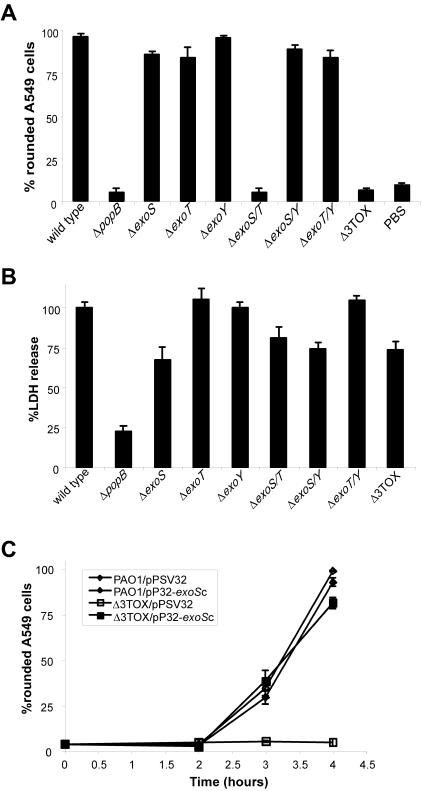
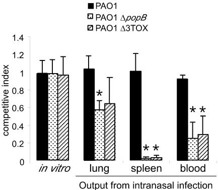
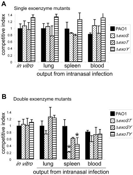
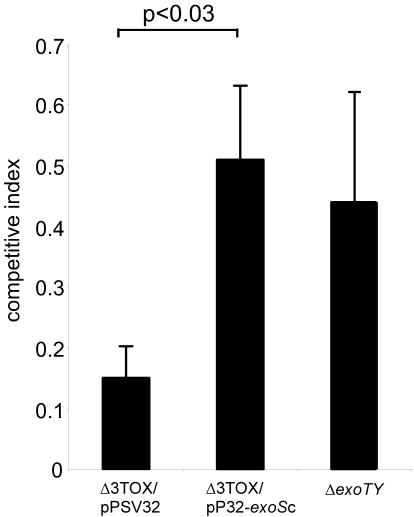
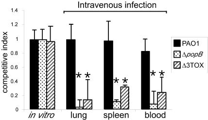
Pseudomonas aeruginosa uses a dedicated type III secretion system to deliver toxins directly into the cytoplasm of host cells. While progress has been made in elucidating the function of type III-secreted toxins in vitro, the in vivo functions of the type III-secreted exoenzymes are less well understood, particularly for the sequenced strain PAO1. Therefore, we have systematically deleted the genes for the three known type III effector molecules (exoS, exoT, and exoY) in P. aeruginosa PAO1 and assayed the effect of the deletions, both singly and in combination, on cytotoxicity in vitro and in vivo. We found that the type III secretion system acts differently on different cell types, causing an exoST-dependent rounding of a lung epithelial-like cell line in contrast to causing an exoSTY-independent but translocase (popB)-dependent lysis of a macrophage cell line. We utilized an in vivo competitive infection model to test each of our mutants, examining replication in the lung and spread to secondary sites such as the blood and spleen. Type III mutants inoculated intranasally exhibited only a minor defect in replication and survival in the lung, but popB and exoSTY triple mutants were profoundly defective in their ability to spread systemically. Intravenous injection of the mutants indicated that the type III secretion machinery is required for survival in the blood. Furthermore, our findings suggest that the effector-independent popB-dependent cytotoxicity that we and others have observed in vitro in macrophage cell lines may not be of great importance in vivo.


Figures

FIG. 1.
FIG. 1.
Differential effects of the type III secretion system on cultured cells in vitro. (A) Rounding of A549 lung epithelial cell-like cells requires exoenzymes S and T. Cells were exposed to wild-type P. aeruginosa PAO1 or the indicated PAO1 mutant strains for 4 h before fixation and enumeration of the percentage of cells that had rounded up. Δ3TOX is a PAO1 ΔexoS ΔexoT ΔexoY triple mutant. PBS, phosphate-buffered saline. (B) Lysis of RAW264.7 macrophages requires an intact type III secretion system but does not require any of the known secreted effector proteins. The amounts of LDH released from cells ∼5 h after exposure to wild-type P. aeruginosa PAO1 or the indicated PAO1 mutant strains are shown. The amount of enzyme released by detergent was set as 100%, and the amount of enzyme released in mock-treated cells was set as 0%. (C) The cell rounding phenotype of the Δ3TOX mutant can be complemented by expression of exoS from a plasmid. Wild-type PAO1 or PAO1 Δ3TOX was transformed with an ExoS expression plasmid (pP32-exoSc) or an empty vector control plasmid (pPSV32), and the extent of A549 cell rounding was assessed at various time points after infection.
FIG. 2.
FIG. 2.
Type III secretion mutants administered intranasally exhibit a mild defect in replication in the lung but exhibit reduced spread and survival in secondary sites, such as spleen and blood. Wild-type P. aeruginosa PAO1 or isogenic ΔpopB or Δ3TOX mutants (producing white colonies on X-Gal plates) were mixed 1:1 with wild-type P. aeruginosa PAO1 carrying lacZ at a neutral phage attachment site (attB; produces blue colonies on X-Gal plates). The CI was calculated as the ratio of white to blue colonies in the output sample divided by the ratio of white to blue colonies in the input sample. In vitro-grown samples consisted of a 1:1,000 dilution of the inoculum grown overnight in Luria broth. Δ3TOX is a PAO1 ΔexoS ΔexoT ΔexoY triple mutant. At least three mice were analyzed for each genotype. *, P < 0.02 versus wild-type PAO1 for each tissue.
FIG. 3.
FIG. 3.
Systematic analysis of in vivo growth and survival of single and double exoenzyme mutants. (A) PAO1 and isogenic mutants carrying in-frame deletions of exoenzyme S, T, or Y were inoculated intranasally (or grown in vitro) in competition with PAO1 attB::lacZ. After 21 h, the indicated tissues were homogenized and plated on X-Gal plates and the ratio of white to blue colonies was enumerated. The CI was calculated as the ratio of white to blue colonies in the output sample divided by the ratio of white to blue colonies in the input sample. At least three mice were analyzed for each genotype. (B) Same as panel A, but double mutants were tested. Data from Fig. 2 for PAO1 are shown for comparison. *, P < 0.02 versus wild-type PAO1 for each tissue.
FIG. 4.
FIG. 4.
An exoS expression plasmid can complement the growth and survival defect of a Δ3TOX (ΔexoS ΔexoT ΔexoY) mutant. PAO1 Δ3TOX was transformed with a plasmid expressing exoS (pP32-exoSc) or with an empty vector control plasmid (pPSV32). The two strains were inoculated intranasally, and the CI for the spleen was calculated as for Fig. 2. The testing of individual recovered colonies confirmed that both plasmids were stably maintained during in vivo growth. The CI for the ΔexoTY mutant (from Fig. 3) is shown for comparison. Student's t test was used to calculate P values.
FIG. 5.
FIG. 5.
The type III secretion system is required for survival of P. aeruginosa in the blood. The indicated mutant strains were inoculated intravenously, and 21 h later lungs, spleens, and blood were recovered from infected animals. The CI was calculated as for Fig. 2. *, P < 0.02 versus wild-type PAO1 for each tissue.

Similar articles

Cited by

References

    1. Barbieri, A. M., Q. Sha, P. Bette-Bobillo, P. D. Stahl, and M. Vidal. 2001. ADP-ribosylation of Rab5 by ExoS of Pseudomonas aeruginosa affects endocytosis. Infect. Immun. 69:5329-5334. - PMC - PubMed
    1. Barbieri, J. T. 2000. Pseudomonas aeruginosa exoenzyme S, a bifunctional type-III secreted cytotoxin. Int. J. Med. Microbiol. 290:381-387. - PubMed
    1. Becher, A., and H. P. Schweizer. 2000. Integration-proficient Pseudomonas aeruginosa vectors for isolation of single-copy chromosomal lacZ and lux gene fusions. BioTechniques 29:948-950, 952. - PubMed
    1. Coburn, J., and D. W. Frank. 1999. Macrophages and epithelial cells respond differently to the Pseudomonas aeruginosa type III secretion system. Infect. Immun. 67:3151-3154. - PMC - PubMed
    1. Dacheux, D., J. Goure, J. Chabert, Y. Usson, and I. Attree. 2001. Pore-forming activity of type III system-secreted proteins leads to oncosis of Pseudomonas aeruginosa-infected macrophages. Mol. Microbiol. 40:76-85. - PubMed

Publication types

MeSH terms